

")


Диагностика аневризм грудного отдела аорты и патологического расширения лёгочного ствола с использованием компьютерной томографии органов грудной клетки и искусственного интеллекта: современные подходы и перспективы (научный обзор)
- Авторы: Соловьёв А.В.1,2, Синицын В.Е.1,3, Владзимирский А.В.1, Памова А.П.1
-
Учреждения:
- Научно-практический клинический центр диагностики и телемедицинских технологий
- Морозовская детская городская клиническая больница
- Московский государственный университет имени М.В. Ломоносова
- Выпуск: Том 6, № 2 (2025)
- Страницы: 286-301
- Раздел: Обзоры
- URL: https://journals.rcsi.science/DD/article/view/310216
- DOI: https://doi.org/10.17816/DD641679
- EDN: https://elibrary.ru/QHBRWF
- ID: 310216
Цитировать
Полный текст

Аннотация
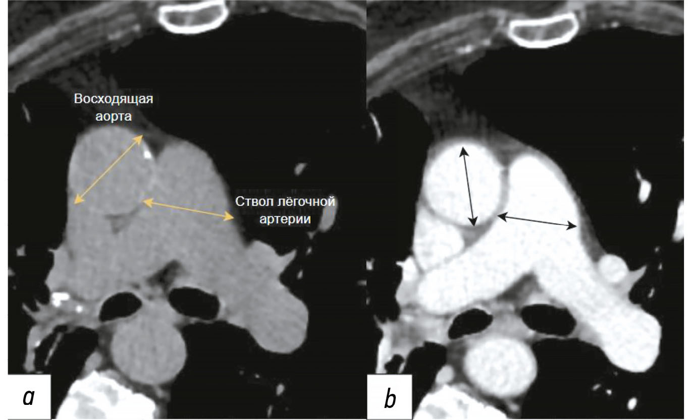
Ранняя диагностика аневризм грудного отдела аорты и патологического расширения лёгочного ствола имеет решающее значение для предотвращения серьёзных осложнений, включая разрыв сосудистой стенки и острую правожелудочковую недостаточность, а также для снижения смертности от сердечно-сосудистых заболеваний. В представленном обзоре рассматриваются современные подходы к визуализации этих патологий, с акцентом на использование компьютерной томографии в качестве «золотого стандарта». Отдельное внимание уделено внедрению технологий искусственного интеллекта, которые позволяют автоматически сегментировать сосудистые структуры, измерять их диаметр и проводить оппортунистический скрининг, выявляя скрытые патологии на ранних стадиях без необходимости проведения дополнительных исследований, что снижает нагрузку на врачей-рентгенологов и повышает качество медицинской помощи. Подробно анализируется опыт Московского эксперимента, в рамках которого использование технологий искусственного интеллекта в анализе медицинских изображений показало высокую чувствительность, воспроизводимость и сокращение времени описания. Несмотря на значительные преимущества, подчёркивается необходимость контроля результатов работы искусственного интеллекта специалистами для обеспечения точности и надёжности диагностики. Также отмечается актуальность адаптации алгоритмов к разным протоколам сканирования и популяционным особенностям. Кроме того, подчёркивается важность междисциплинарного взаимодействия кардиологов, рентгенологов, инженеров-данных и разработчиков программного обеспечения для для эффективного внедрения в рутинную клиническую деятельность. В заключение делается вывод о значительном потенциале технологий искусственного интеллекта для повышения качества диагностики и подчёркивается необходимость дальнейших клинических исследований и стандартизации методик для их успешной интеграции в повседневную практику.
Полный текст
Открыть статью на сайте журналаОб авторах
Александр Владимирович Соловьёв
Научно-практический клинический центр диагностики и телемедицинских технологий; Морозовская детская городская клиническая больница
Автор, ответственный за переписку.
Email: atlantis.92@mail.ru
ORCID iD: 0000-0003-4485-2638
SPIN-код: 9654-4005
MD
Россия, Москва; МоскваВалентин Евгеньевич Синицын
Научно-практический клинический центр диагностики и телемедицинских технологий; Московский государственный университет имени М.В. Ломоносова
Email: vsini@mail.ru
ORCID iD: 0000-0002-5649-2193
SPIN-код: 8449-6590
д-р мед. наук, профессор
Россия, Москва; МоскваАнтон Вячеславович Владзимирский
Научно-практический клинический центр диагностики и телемедицинских технологий
Email: VladzimirskijAV@zdrav.mos.ru
ORCID iD: 0000-0002-2990-7736
SPIN-код: 3602-7120
д-р мед. наук
Россия, МоскваАнастасия Петровна Памова
Научно-практический клинический центр диагностики и телемедицинских технологий
Email: PamovaAP@zdrav.mos.ru
ORCID iD: 0000-0002-0041-3281
SPIN-код: 5146-4355
канд. мед. наук
Россия, МоскваСписок литературы
- Wang X, Zhu H. Artificial intelligence in image-based cardiovascular disease analysis: a comprehensive survey and future outlook. Journal of Latex Class Files. 2024;14(8):1–22. doi: 10.48550/arXiv.2402.03394
- Nagibina YuV, Zakharova LA. Life quality, medical and social characteristics of coronary heart disease patients. Russian Journal of Cardiology. 2017;22(3):155–159. doi: 10.15829/1560-4071-2017-3-155-159 EDN: YHOFDL
- Czerny M, Grabenwöger M, Berger T, et al. EACTS/STS guidelines for diagnosing and treating acute and chronic syndromes of the aortic organ. The Annals of Thoracic Surgery. 2024;118(1):5–115. doi: 10.1016/j.athoracsur.2024.01.021 EDN: XLPIEC
- Chernina VYu, Blohin IA, Nikolaev AE, et al. Tactics of incidentaloma management. Section 3. Thyroid gland, pituitary gland, vessels and mediastinum: methodical recommendations. Moscow: Research and Practical Clinical Center for Diagnostics and Telemedicine Technologies; 2019. (In Russ.) [cited 2024 May 12]. Available from: https://niioz.ru/upload/iblock/62c/62ceeb66d4528c831ddf40ea3f918d01.pdf
- Erbel R, Aboyans V, Boileau C, et al.; ESC Committee for Practice Guidelines. 2014 ESC Guidelines on the diagnosis and treatment of aortic diseases: Document covering acute and chronic aortic diseases of the thoracic and abdominal aorta of the adult. The Task Force for the Diagnosis and Treatment of Aortic Diseases of the European. Eur. Heart J. 2014;35(41):2873–2926. doi: 10.1093/eurheartj/ehu281
- Gouveia E Melo R, Silva Duarte G, Lopes A, et al. Incidence and prevalence of thoracic aortic aneurysms: a systematic review and meta-analysis of population-based studies. Semin. Thorac. Cardiovasc. Surg. 2022;34(1):1–16. doi: 10.1053/j.semtcvs.2021.02.029
- Abugov SA, Averina TB, Akchurin RS, et al. Clinical guidelines. guidelines for the diagnosis and treatment of aortic diseases (2017). Russian Journal Of Cardiology And Cardiovascular Surgery. 2018;11(1):7–67. EDN: YPAKRP
- Galiè N, Humbert M, Vachiery JL, et al. 2015 ESC/ERS Guidelines for the diagnosis and treatment of pulmonary hypertension. European Respiratory Journal. 2015;46(4):903–975. doi: 10.1183/13993003.01032-2015 EDN: XYRISJ
- Humbert M, Kovacs G, Hoeper MM, et al; the ESC/ERS scientific document group. 2022 ESC/ERS Guidelines for the diagnosis and treatment of pulmonary hypertension. European Respiratory Journal. 2022;61(1):2200879. doi: 10.1183/13993003.00879-2022 EDN: DMBOSS
- Chazova IE, Martynyuk TV, Valieva ZS, et al. Eurasian clinical guidelines on diagnosis and treatment of pulmonary hypertension. Eurasian heart journal. 2020;(1):78–122. doi: 10.38109/2225-1685-2020-1-78-122 EDN: YOTXVT
- Sampson UKA, Norman PE, Fowkes FGR, et al. Global and regional burden of aortic dissection and aneurysms: mortality trends in 21 world regions, 1990 to 2010. Global Heart. 2014;9(1):171–180. doi: 10.1016/j.gheart.2013.12.010 EDN: SPJFQB
- Sampson UKA, Norman PE, Fowkes FGR, et al. Estimation of global and regional incidence and prevalence of abdominal aortic aneurysms 1990 to 2010. Global Heart. 2014;9(1):159–170. doi: 10.1016/j.gheart.2013.12.009 EDN: SPKAKV
- Krafcik BM, Stone DH, Cai M, et al. Changes in global mortality from aortic aneurysm. Journal of Vascular Surgery. 2024;80(1):81–88.e1. doi: 10.1016/j.jvs.2024.02.025 EDN: ZUUPKL
- Irtyuga OB, Voronkina IV, Smagina LV, et al. Тhe frequency to detect of ascending aorta aneurysms and the mechanism of its development according register of the аlmazov federal heart, blood and endocrinology centre. Almazov Federal Heart, Blood And Endocrinology Centre Bulletin. 2011;(5):73–78. EDN: OWGHOB
- Kuznechevsky F.V., Osipov A.Kh., Evsikov E.M., Abramov I.S., Otarova S.M. Prevalence and clinical features of aorta aneurysm; and dissections: 10-year results of consequent autopsies made at O.M. Filatov City Clinical Hospital №15. Russian Journal of Cardiology. 2004;9(6):5–13. EDN: ISVRYL
- Pradella M, Achermann R, Sperl JI, et al. Performance of a deep learning tool to detect missed aortic dilatation in a large chest CT cohort. Frontiers in Cardiovascular Medicine. 2022;9:972512. doi: 10.3389/fcvm.2022.972512 EDN: NWYDJQ
- Lavall D, Schäfers HJ, Böhm M, Laufs U. Aneurysms of the ascending aorta. Deutsches Ärzteblatt international. 2012;109(13):227–233. doi: 10.3238/arztebl.2012.0227
- Elefteriades JA. Natural history of thoracic aortic aneurysms: indications for surgery, and surgical versus nonsurgical risks. The Annals of Thoracic Surgery. 2002;74(5):S1877–S1880. doi: 10.1016/S0003-4975(02)04147-4
- Wong RHL, Yang F, Fujikawa T, et al. Pocket-size mobile echocardiographic screening of thoracic aortic aneurysms in hypertensive patients. The Annals of Thoracic Surgery. 2021;111(5):1554–1559. doi: 10.1016/j.athoracsur.2020.07.018 EDN: IRQRDZ
- Lum RTW, Ho JYK, Chow SCY, et al. Screening for thoracic aortic aneurysm. Journal of the Hong Kong College of Cardiology. 2024;31(4). doi: 10.55503/2790-6744.1538 EDN: YKXGBT
- McClure RS, Brogly SB, Lajkosz K, et al. Epidemiology and management of thoracic aortic dissections and thoracic aortic aneurysms in Ontario, Canada: A population-based study. The Journal of Thoracic and Cardiovascular Surgery. 2018;155(6):2254–2264.e4. doi: 10.1016/j.jtcvs.2017.11.105
- Olsson C, Thelin S, Ståhle E, et al. Thoracic aortic aneurysm and dissection. Circulation. 2006;114(24):2611–2618. doi: 10.1161/CIRCULATIONAHA.106.630400
- Cho IJ, Jang SY, Chang HJ, et al. Aortic Aneurysm screening in a high-risk population: a non-contrast computed tomography study in korean males with hypertension. Korean Circulation Journal. 2014;44(3):162–169. doi: 10.4070/kcj.2014.44.3.162
- Kato K, Oguri M, Kato N, et al. Assessment of genetic risk factors for thoracic aortic aneurysm in hypertensive patients. American Journal of Hypertension. 2008;21(9):1023–1027. doi: 10.1038/ajh.2008.229
- Mehrabi Nasab E, Athari SS. The prevalence of thoracic aorta aneurysm as an important cardiovascular disease in the general population. Journal of Cardiothoracic Surgery. 2022;17(1):1–6. doi: 10.1186/s13019-022-01767-0 EDN: QOWOWT
- Vandroux D, Aboyans V, Houehanou YC, et al; TAHES study investigators. Normal values of proximal aorta diameters in healthy Sub Saharan Africans: The TAHES study. Echocardiography. 2022;39(4):576–583. doi: 10.1111/echo.15331 EDN: TGBSWV
- Clouse WD, Hallett, Jr JW, Schaff HV, et al. Improved prognosis of thoracic aortic aneurysms. JAMA. 1998;280(22):1926–1929. doi: 10.1001/jama.280.22.1926
- Elefteriades JA. Thoracic aortic aneurysm: reading ttable he enemy’s playbook. World Journal of Surgery. 2008;32(3):366–374. doi: 10.1007/s00268-007-9398-3 EDN: EAAZYC
- Miller WT. Thoracic aortic aneurysms: plain film findings. Seminars in Roentgenology. 2001;36(4):288–294. doi: 10.1053/sroe.2001.26937
- von Kodolitsch Y, Nienaber CA, Dieckmann C, et al. Chest radiography for the diagnosis of acute aortic syndrome. The American Journal of Medicine. 2004;116(2):73–77. doi: 10.1016/j.amjmed.2003.08.030
- Flachskampf FA, Badano L, Daniel WG, et al; for the European Association of Echocardiography; endorsed by the Echo Committee of the European Association of Cardiothoracic Anaesthesiologists. Recommendations for transoesophageal echocardiography: update 2010. European Journal of Echocardiography. 2010;11(7):557–576. doi: 10.1093/ejechocard/jeq057
- le Polain de Waroux JB, Pouleur AC, Goffinet C, et al. Functional anatomy of aortic regurgitation: accuracy, prediction of surgical repairability, and outcome implications of transesophageal echocardiography. Circulation. 2007;116(Suppl. 11):I264–I269. doi: 10.1161/CIRCULATIONAHA.106.680074
- Kallianos KG, Burris NS. Imaging thoracic aortic aneurysm. Radiologic Clinics of North America. 2020;58(4):721–731. doi: 10.1016/j.rcl.2020.02.009 EDN: CAUYQG
- Lau C, Feldman DN, Girardi LN, Kim LK. Imaging for surveillance and operative management for endovascular aortic aneurysm repairs. Journal of Thoracic Disease. 2017;9(S4):S309–S316. doi: 10.21037/jtd.2017.03.89
- Chiu KWH, Ling L, Tripathi V, et al. Ultrasound measurement for abdominal aortic aneurysm screening: a direct comparison of the three leading methods. European Journal of Vascular and Endovascular Surgery. 2014;47(4):367–373. doi: 10.1016/j.ejvs.2013.12.026
- Manning BJ, Kristmundsson T, Sonesson B, Resch T. Abdominal aortic aneurysm diameter: a comparison of ultrasound measurements with those from standard and three-dimensional computed tomography reconstruction. Journal of Vascular Surgery. 2009;50(2):263–268. doi: 10.1016/j.jvs.2009.02.243
- Lansac E, Di Centa I, Raoux F, et al. A lesional classification to standardize surgical management of aortic insufficiency towards valve repair. European Journal of Cardio-Thoracic Surgery. 2008;33(5):872–878. doi: 10.1016/j.ejcts.2007.12.033
- Rybakova MK, Alehin MN, Mitkov VV. Practical guide to ultrasound diagnostics. Echocardiography. Moscow: Vidar-M; 2008. (In Russ.) ISBN: 978-5-88429-118-8 [cited 2024 May 12]. Available from: http://vidar.ru/BookImg/001-020_Rybakova.pdf
- Roelandt JR, Bruining N, Bom N. Perspectives in cardiac ultrasound. Przeglad Lekarski. 2002;59(8):557–561.
- Gavrilenkov VI, Kuznecov AA, Perlej VE, et al. Echocardiographic assessment of normal aortic valve biomechanics. Ultrasound And Functional Diagnostics. 2003;(2):89–96. (In Russ.)
- Claridge R, Arnold S, Morrison N, van Rij AM. Measuring abdominal aortic diameters in routine abdominal computed tomography scans and implications for abdominal aortic aneurysm screening. Journal of Vascular Surgery. 2017;65(6):1637–1642. doi: 10.1016/j.jvs.2016.11.044
- Hall T, Shah P, Wahi S. The role of transesophageal echocardiography in aortic valve preserving procedures. Indian Heart Journal. 2014;66(3):327–333. doi: 10.1016/j.ihj.2014.05.001
- Downing SW, Sperling JS, Mirvis SE, et al. Experience with spiral computed tomography as the sole diagnostic method for traumatic aortic rupture. The Annals of Thoracic Surgery. 2001;72(2):495–502. doi: 10.1016/S0003-4975(01)02827-2
- Rémy-Jardin M, Bonnel F, Masson P, et al. Outil optimal de dépistage en pathologie thoracique: radiographie ou scanner? J Radiol. 2001;82(9 Pt 2):1108–1118. (In French)
- Ellis JD, Mayo JR. Computed tomography evaluation of traumatic rupture of the thoracic aorta: an outcome study. Can Assoc Radiol J. 2007;58(1):22–26.
- Szymczyk K, Polguj M, Szymczyk E, et al. Assessment of aortic valve in regard to its anatomical variants morphology in 2053 patients using 64-slice CT retrospective coronary angiography. BMC Cardiovascular Disorders. 2016;16(1):1–9. doi: 10.1186/s12872-016-0261-z EDN: RPMNFG
- Lee HY, Kim SM, Lee KS, et al. Quantification of Aortic Valve Calcifications Detected During Lung Cancer-Screening CT Helps Stratify Subjects Necessitating Echocardiography for Aortic Stenosis Diagnosis. Medicine. 2016;95(19):e3710. doi: 10.1097/MD.0000000000003710
- Belov YuV, Kertes MI, Bogopolskaya OM, et al. Strategy and tactics of instrumental examination of patients with thoracic and thoracoabdominal aortic aneurysm. Angiology and Vascular Surgery. Journal Named After Academician A.V. Pokrovsky. 2005;11(4):33–51. EDN: PFRPXT
- Tops LF, Wood DA, Delgado V, et al. Noninvasive evaluation of the aortic root with multislice computed tomography. JACC: Cardiovascular Imaging. 2008;1(3):321–330. doi: 10.1016/j.jcmg.2007.12.006
- Weinreb JC, Rodby RA, Yee J, et al. Use of intravenous gadolinium-based contrast media in patients with kidney disease: consensus statements from the American College of Radiology and the National Kidney Foundation. Radiology. 2021;298(1):28–35. doi: 10.1148/radiol.2020202903 EDN: WZBRMI
- Hagan PG, Nienaber CA, Isselbacher EM, et al. The International Registry of Acute Aortic Dissection (IRAD). JAMA. 2000;283(7):897–903. doi: 10.1001/jama.283.7.897
- Bokeria LA, Malashenkov AL, Makarenko VN, et al. Spiral computed tomographyin aortic aneurysm diagnostics. Annals of the Russian Academy of Medical Sciences. 2005;(4):25–31. EDN: HRXFHZ
- Prozorov SA, Belozerov GE, Dubrov EYa, et al. diagnostic radiology of multiple aortic aneurysms. Medical Visualization. 2005;(3):83–87. EDN: TGLBBB
- Sommer T, Fehske W, Holzknecht N, et al. Aortic dissection: a comparative study of diagnosis with spiral CT, multiplanar transesophageal echocardiography, and MR imaging. Radiology. 1996;199(2):347–352. doi: 10.1148/radiology.199.2.8668776
- Kapustin AJ, Litt HI. Diagnostic imaging for aortic dissection. Seminars in Thoracic and Cardiovascular Surgery. 2005;17(3):214–223. doi: 10.1053/j.semtcvs.2005.06.006
- Tsai TT, Nienaber CA, Eagle KA. Acute aortic syndromes. Circulation. 2005;112(24):3802–3813. doi: 10.1161/CIRCULATIONAHA.105.534198
- Cigarroa JE, Isselbacher EM, DeSanctis RW, Eagle KA. Diagnostic imaging in the evaluation of suspected aortic dissection — old standards and new directions. New England Journal of Medicine. 1993;328(1):35–43. doi: 10.1056/NEJM199301073280107
- Tyutin LA, Yakovleva EK. Magnetic resonance angiography: development stages, diagnostic potential and limitations. Medical Visualization. 2013;(2):29–40. EDN: RBJGKL
- François CJ, Carr JC. MRI of the thoracic aorta. Magnetic Resonance Imaging Clinics Of North America. 2007;15(4):639–651. doi: 10.1016/j.mric.2007.08.011
- Nienaber CA. The role of imaging in acute aortic syndromes. European Heart Journal — Cardiovascular Imaging. 2012;14(1):15–23. doi: 10.1093/ehjci/jes215
- Litmanovich D, Bankier AA, Cantin L, et al. CT and MRI in Diseases of the Aorta. American Journal of Roentgenology. 2009;193(4):928–940. doi: 10.2214/AJR.08.2166
- Barker AJ, Markl M, Bürk J, et al. Bicuspid Aortic Valve Is Associated With Altered Wall Shear Stress in the Ascending Aorta. Circulation: Cardiovascular Imaging. 2012;5(4):457–466. doi: 10.1161/CIRCIMAGING.112.973370
- Blockmans D, Ceuninck L, Vanderschueren S, et al. Repetitive 18F-fluorodeoxyglucose positron emission tomography in giant cell arteritis: a prospective study of 35 patients. Arthritis Care & Research. 2006;55(1):131–137. doi: 10.1002/art.21699
- Walter MA, Melzer RA, Schindler C, et al. The value of [18F]FDG-PET in the diagnosis of large-vessel vasculitis and the assessment of activity and extent of disease. European Journal of Nuclear Medicine and Molecular Imaging. 2005;32(6):674–681. doi: 10.1007/s00259-004-1757-9 EDN: QAVZJE
- Kuehl H, Eggebrecht H, Boes T, et al. Detection of inflammation in patients with acute aortic syndrome: comparison of FDG-PET/CT imaging and serological markers of inflammation. Heart. 2008;94(11):1472–1477. doi: 10.1136/hrt.2007.127282
- Tokuda Y, Oshima H, Araki Y, et al. Detection of thoracic aortic prosthetic graft infection with 18F-fluorodeoxyglucose positron emission tomography/computed tomography. European Journal of Cardio-Thoracic Surgery. 2013;43(6):1183–1187. doi: 10.1093/ejcts/ezs693
- Fiorucci B, Banafsche R, Jerkku T, et al. Das thorakale Aortenaneurysma — Diagnostik und Behandlungsstrategien. Deutsche medizinische Wochenschrift (1946). 2019;144(3):146–151. (In German) doi: 10.1055/a-0648-0207
- Feldman L. Digital Subtraction Angiography of the Chest. Clinics in Chest Medicine. 1984;5(2):313–328. doi: 10.1016/S0272-5231(21)00254-9
- Rauber K, Kollath J. Die Diagnose der Aortenruptur durch digitale Subtraktionsangiographie (DSA). RoFo: Fortschritte auf dem Gebiete der Rontgenstrahlen und der Nuklearmedizin. 1983;139(2):167–170. (In German) doi: 10.1055/s-2008-1055864
- Lambelin M, Janssens L, Haenen L. Iatrogenic ascending aorta dissection during diagnostic coronary angiography: rare but life-threatening. Case Reports in Cardiology. 2014;2014:1–3. doi: 10.1155/2014/809398
- Carpenter SW, Kodolitsch YV, Debus ES, et al. Acute aortic syndromes: definition, prognosis and treatment options. The Journal of Cardiovascular Surgery. 2014;55(2 Suppl 1):133–144.
- Hannuksela M, Lundqvist S, Carlberg B. Thoracic aorta — dilated or not? Scandinavian Cardiovascular Journal. 2006;40(3):175–178. doi: 10.1080/14017430600565999
- Davies RR, Gallo A, Coady MA, et al. Novel measurement of relative aortic size predicts rupture of thoracic aortic aneurysms. The Annals of Thoracic Surgery. 2006;81(1):169–177. doi: 10.1016/j.athoracsur.2005.06.026
- Girardi LN, Lau C, Gambardella I. Aortic dimensions as predictors of adverse events. The Journal of Thoracic and Cardiovascular Surgery. 2021;161(4):1193–1197. doi: 10.1016/j.jtcvs.2020.06.137 EDN: GNERDK
- Hoeper MM, Humbert M, Souza R, et al. A global view of pulmonary hypertension. The Lancet Respiratory Medicine. 2016;4(4):306–322. doi: 10.1016/S2213-2600(15)00543-3 EDN: WUFYUX
- Naing P, Kangaharan N, Scalia GM, et al. Pulmonary hypertension in remote and disadvantaged population: overcoming unique challenges for improved outcomes. Internal Medicine Journal. 2022;53(1):12–20. doi: 10.1111/imj.15860 EDN: YNYRXI
- Martynjuk TV. Pulmonary hypertension: diagnosis and treatment. Moscow: Publishing House “Medical Information Agency”; 2018. (In Russ.) EDN: UXDPPX
- Aliev AF, Kudryavtsev ND, Petraikin AV, et al. Changing of pulmonary artery diameter in accordance with severity of COVID-19 (assessment based on non-contrast computer tomography). Digital Diagnostics. 2021;2(3):249–260. doi: 10.17816/DD76726 EDN: VTMKCJ
- Chuchalin A, Khaltaev N, Antonov N, et al. Chronic respiratory diseases and risk factors in 12 regions of the Russian Federation. International Journal of Chronic Obstructive Pulmonary Disease. 2014;9:963–974. doi: 10.2147/COPD.S67283 EDN: UEYOFH
- Avdeev SN, Ajsanov ZR, Belevskij AS, et al; Russian Respiratory Society. Clinical guidelines: chronic obstructive pulmonary disease. 2nd ed, revised and supplemented. Moscow: Atmosfera; 2007. (In Russ.) EDN: QLPFTN
- Galiè N, Simonneau G. The fifth world symposium on pulmonary hypertension. Journal of the American College of Cardiology. 2013;62(25):D1–D3. doi: 10.1016/j.jacc.2013.10.030 EDN: YEGEFV
- Badesch DB, Raskob GE, Elliott CG, et al. Pulmonary arterial hypertension. Chest. 2010;137(2):376–387. doi: 10.1378/chest.09-1140
- Hoeper MM, Bogaard HJ, Condliffe R, et al. Definitions and diagnosis of pulmonary hypertension. Journal of the American College of Cardiology. 2013;62(25):D42–D50. doi: 10.1016/j.jacc.2013.10.032
- Brown LM, Chen H, Halpern S, et al. Delay in recognition of pulmonary arterial hypertension. Chest. 2011;140(1):19–26. doi: 10.1378/chest.10-1166
- Tonelli AR, Ascha M, Renapurkar RD. A review of imaging modalities in pulmonary hypertension. Annals of Thoracic Medicine. 2017;12(2):61–73. doi: 10.4103/1817-1737.203742 EDN: YDBKBN
- Avdeev SN, Barbarash OL, Valieva ZS, et al. 2024 Clinical practice guidelines for Pulmonary hypertension, including chronic thromboembolic pulmonary hypertension. Russian Journal of Cardiology. 2024;29(11):170–250. doi: 10.15829/1560-4071-2024-6161 EDN: MYEOVA
- Zouk AN, Gulati S, Xing D, et al. Pulmonary artery enlargement is associated with pulmonary hypertension and decreased survival in severe cystic fibrosis: a cohort study. PLOS ONE. 2020;15(2):e0229173. doi: 10.1371/journal.pone.0229173 EDN: FJNDMS
- Dwivedi K, Sharkey M, Condliffe R, et al. Pulmonary hypertension in association with lung disease: quantitative CT and artificial intelligence to the rescue? State-of-the-Art Review. Diagnostics. 2021;11(4):679. doi: 10.3390/diagnostics11040679 EDN: QCTQAF
- Truong QA, Massaro JM, Rogers IS, et al. Reference values for normal pulmonary artery dimensions by noncontrast cardiac computed tomography. Circulation: Cardiovascular Imaging. 2012;5(1):147–154. doi: 10.1161/CIRCIMAGING.111.968610
- Lewis G, Hoey ET, Reynolds JH, et al. Multi-detector CT assessment in pulmonary hypertension: techniques, systematic approach to interpretation and key findings. Quantitative imaging in Medicine and Surgery. 2015;5(3):423–432. doi: 10.3978/j.issn.2223-4292.2015.01.05
- Nakanishi R, Rana JS, Shalev A, et al. mortality risk as a function of the ratio of pulmonary trunk to ascending aorta diameter in patients with suspected coronary artery disease. The American Journal of Cardiology. 2013;111(9):1259–1263. doi: 10.1016/j.amjcard.2013.01.266
- Lee SH, Kim YJ, Lee HJ, et al. Comparison of CT-determined pulmonary artery diameter, aortic diameter, and their ratio in healthy and diverse clinical conditions. PLOS ONE. 2015;10(5):e0126646. doi: 10.1371/journal.pone.0126646
- Raju SN, Pandey NN, Sharma A, et al. Pulmonary Arterial Dilatation: Imaging Evaluation Using Multidetector Computed Tomography. Indian Journal of Radiology and Imaging. 2021;31(2):409–420. doi: 10.1055/s-0041-1734225 EDN: NEUMWO
- Spagnolo P, Cozzi A, Foà RA, et al. CT-derived pulmonary vascular metrics and clinical outcome in COVID-19 patients. Quantitative Imaging in Medicine and Surgery. 2020;10(6):1325–1333. doi: 10.21037/QIMS-20-546 EDN: WIOGYX
- Markl M, Hope MD. 4D flow imaging—state of the art. Annals of Cardiothoracic Surgery. 2022;11(4):468–469. doi: 10.21037/acs-2021-bav-15 EDN: YIIRBV
- Law M. “Opportunistic” screening. Journal of Medical Screening. 1994;1(4):208. doi: 10.1177/096914139400100403
- Solovev AV, Vasilev YuA, Sinitsyn VE, et al. Improving aortic aneurysm detection with artificial intelligence based on chest computed tomography data. Digital Diagnostics. 2024;5(1):29–40. doi: 10.17816/DD569388 EDN: CZNZYP
- Vasan RS. Biomarkers of cardiovascular disease. Circulation. 2006;113(19):2335–2362. doi: 10.1161/CIRCULATIONAHA.104.482570 EDN: MFJKKD
- Urbanowicz T, Rajewska-Tabor J, Olasińska-Wiśniewska A, et al. Demographical and clinical factors predictive for aortic dilatation. When should we be concerned about the size? Reviews in Cardiovascular Medicine. 2024;25(5):150. doi: 10.31083/j.rcm2505150 EDN: DIDZNL
- Mintz Y, Brodie R. Introduction to artificial intelligence in medicine. Minimally Invasive Therapy & Allied Technologies. 2019;28(2):73–81. doi: 10.1080/13645706.2019.1575882 EDN: EWABMW
- Goncharov M, Pisov M, Shevtsov A, et al. CT-Based COVID-19 triage: deep multitask learning improves joint identification and severity quantification. Medical Image Analysis. 2021;71:102054. doi: 10.1016/j.media.2021.102054 EDN: RAHCWT
- Vladzymyrsky AV, Vasilev YuA, Arzamasov KM, et al. Computer vision in radiation diagnostics: the first stage of the Moscow experiment. 2nd ed. Moscow: Izdatel'skie resheniya; 2023 (In Russ.) ISBN: 978-5-0059-3043-9 EDN: FOYLXK
- Vasilev YA, Bobrovskaya TM, Arzamasov KM, et al. Medical datasets for machine learning: fundamental principles of standartization and systematization. Manager Zdravookhranenia. 2023;(4):28–41. doi: 10.21045/1811-0185-2023-4-28-41 EDN: EPGAMD
- Petraikin AV, Artyukova ZR, Nisovtsova LA, et al. Analysis of the effectiveness of implementing screening of osteoporosis. Manager Zdravoochranenia. 2021;(2):31–39. doi: 10.21045/1811-0185-2021-2-31-39 EDN: AVDSIW
- Mets OM, de Jong PA, Prokop M. Computed tomographic screening for lung cancer. JAMA. 2012;308(14):1433–1434. doi: 10.1001/jama.2012.12656
- Patel K, Zafar MA, Ziganshin BA, Elefteriades JA. Diabetes mellitus: is it protective against aneurysm? A narrative review. Cardiology. 2018;141(2):107–122. doi: 10.1159/000490373 EDN: IODHHA
- Song L, Zhao S, Wang L, et al. Cardiovascular changes in patients with COVID-19 from Wuhan, China. Frontiers in Cardiovascular Medicine. 2020;7:566484. doi: 10.3389/fcvm.2020.00150 EDN: UNZBRV
- Ostberg N, Zafar M, Ziganshin B, Elefteriades J. The genetics of thoracic aortic aneurysms and dissection: a clinical perspective. Biomolecules. 2020;10(2):182. doi: 10.3390/biom10020182 EDN: XHXLLH
- Eltorai AEM, McKinney SE, Rockenbach MABC, et al. Primary care provider perspectives on the value of opportunistic CT screening. Clinical Imaging. 2024;112:110210. doi: 10.1016/j.clinimag.2024.110210 EDN: MZTJJQ
- Kodenko MR, Vasilev YA, Vladzymyrskyy AV, et al. Diagnostic accuracy of AI for opportunistic screening of abdominal aortic aneurysm in CT: a systematic review and narrative synthesis. Diagnostics. 2022;12(12):3197. doi: 10.3390/diagnostics12123197 EDN: ERWYPX
- Mori M, Bin Mahmood SU, Yousef S, et al. Prevalence of incidentally identified thoracic aortic dilations: insights for screening criteria. Canadian Journal of Cardiology. 2019;35(7):892–898. doi: 10.1016/j.cjca.2019.03.023 EDN: PLXTIZ
- Jiang H, Xu H, Xu Z. Sex-related differences in outcome of thoracic aortic surgery. Journal of Cardiothoracic Surgery. 2024;19(1):1–7. doi: 10.1186/s13019-024-02735-6 EDN: RRSAZK
- Cheung K, Boodhwani M, Chan KL, et al. Thoracic aortic aneurysm growth: role of sex and aneurysm etiology. Journal of the American Heart Association. 2017;6(2):e003792. doi: 10.1161/JAHA.116.003792
- Cayne NS, Veith FJ, Lipsitz EC, et al. Variability of maximal aortic aneurysm diameter measurements on CT scan: significance and methods to minimize. Journal of Vascular Surgery. 2004;39(4):811–815. doi: 10.1016/j.jvs.2003.11.042
- Isselbacher EM. Thoracic and abdominal aortic aneurysms. Circulation. 2005;111(6):816–828. doi: 10.1161/01.CIR.0000154569.08857.7A
- Sedghi Gamechi Z, Bons LR, Giordano M, et al. Automated 3D segmentation and diameter measurement of the thoracic aorta on non-contrast enhanced CT. European Radiology. 2019;29(9):4613–4623. doi: 10.1007/s00330-018-5931-z EDN: TKMBPK
- Monti CB, van Assen M, Stillman AE, et al. Evaluating the performance of a convolutional neural network algorithm for measuring thoracic aortic diameters in a heterogeneous population. Radiology: Artificial Intelligence. 2022;4(2):e210196. doi: 10.1148/ryai.210196 EDN: TJLGGM
- Hamelink II, de Heide EEJ, Pelgrim GJGJ, et al. Validation of an AI-based algorithm for measurement of the thoracic aortic diameter in low-dose chest CT. European Journal of Radiology. 2023;167:111067. doi: 10.1016/j.ejrad.2023.111067
- Kim J, Gupta D, LeComte M, et al. Abstract 17842: 3D visualization and quantitative assessment of the pulmonary arteries on CT using deep learning segmentation. Circulation. 2023;148(Suppl. 1). doi: 10.1161/circ.148.suppl_1.17842 EDN: WREUEP
Дополнительные файлы


