

")


Autoimmune hypophysitis: a case of follow-up during the COVID-19 pandemic period
- Authors: Surovcev E.N.1,2, Zelter P.M.1,3, Kapishnikov A.V.1, Pyshkina Y.S.1
-
Affiliations:
- Samara State Medical University
- Diagnostic and treatment center of International institution for biological systems named after Sergey Berezin
- Meir Hospital
- Issue: Vol 6, No 1 (2025)
- Pages: 178-186
- Section: Case reports
- URL: https://journals.rcsi.science/DD/article/view/310066
- DOI: https://doi.org/10.17816/DD634533
- ID: 310066
Cite item

Abstract
Hypophysitis is a rare inflammatory disorder that affects the pituitary gland and infundibulum, stems from autoimmune, infiltrative, infectious, or unknown causes. Its clinical diagnosis can be challenging because several pituitary lesions, including adenomas and metastases, may clinically present with similar characteristics. Magnetic resonance imaging is crucial for diagnosing suspected cases of hypophysitis and categorizing them as adenohypophysitis (anterior pituitary gland involvement) or infundibulo-neurohypophysitis (pituitary stalk and posterior pituitary involvement). Hypophysitis can be categorized as primary (autoimmune) or secondary due to local lesions (e.g., granulomas, cysts, adenomas) or systemic diseases (e.g., sarcoidosis, Wegener’s granulomatosis). Different factors may have impact on clinical course of hypophysitis. Among them background treatment. These cases have not been sufficiently studied and are practically not presented in publications.
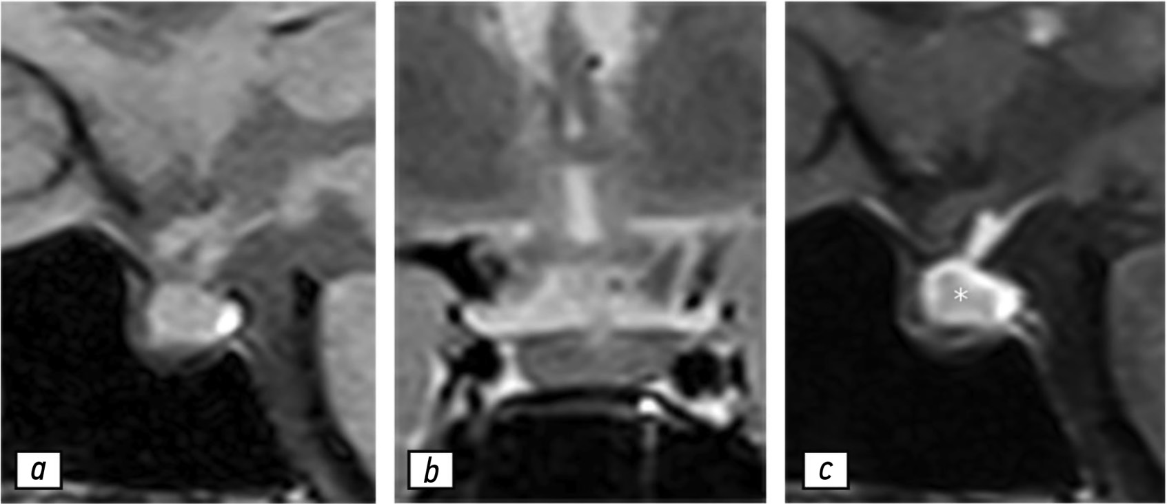
A 37-year-old female with a history of hyperprolactinemia was being treated symptomatically with cabergoline. At first magnetic resonance imaging heterogeneity of the hypophysis was revealed. In September 2021 the follow-up magnetic resonance imaging revealed an increase in the size and heterogeneity of the pituitary gland. In December 2021, the patient developed severe COVID-19-associated pneumonia and was treated with corticosteroids and oxygen support. In May 2022 magnetic resonance imaging revealed a marked increase in the size and heterogeneity of the pituitary gland. Significant clinical and radiological improvement were stated after adding prednisone (10 mg in the morning and 5 mg in the evening) to her treatment.
The patient was followed-up during the COVID-19 pandemic. The management and imaging studies of such patients may be tricky due to the effects related to COVID-19 and its treatment.
During monitoring of hypophysitis, physicians should consider the impact of COVID-19 treatment, particularly corticosteroid therapy, when evaluating the radiological changes.
Full Text
##article.viewOnOriginalSite##About the authors
Evgeniy N. Surovcev
Samara State Medical University; Diagnostic and treatment center of International institution for biological systems named after Sergey Berezin
Email: evgeniisurovcev@mail.ru
ORCID iD: 0000-0002-8236-833X
SPIN-code: 5252-5661
MD, Cand. Sci. (Medicine)
Russian Federation, Samara; TolyattiPavel M. Zelter
Samara State Medical University; Meir Hospital
Email: pzelter@mail.ru
ORCID iD: 0000-0003-1346-5942
SPIN-code: 3678-3932
MD, Cand. Sci. (Medicine)
Russian Federation, Samara; Kfar-Sava, IsraelAleksandr V. Kapishnikov
Samara State Medical University
Email: a.kapishnikov@gmail.com
ORCID iD: 0000-0002-6858-372X
SPIN-code: 6213-7455
MD, Dr. Sci. (Medicine), Professor
Russian Federation, SamaraYuliya S. Pyshkina
Samara State Medical University
Author for correspondence.
Email: yu.pyshkina@yandex.ru
ORCID iD: 0000-0002-7241-6828
SPIN-code: 4225-1020
MD, Cand. Sci. (Medicine), Assistant Professor
Russian Federation, SamaraReferences
- Uccella S, Dottermusch M, Erickson L, et al. Inflammatory and infectious disorders in endocrine pathology. Endocr Pathol. 2023;34(4):406–436. doi: 10.1007/s12022-023-09771-3 EDN: XJTAXG
- Caturegli P. Autoimmune hypophysitis: an underestimated disease in search of its autoantigen(s). J Clin Endocrinol Metab. 2007;92(6):2038–2040. doi: 10.1210/jc.2007-0808
- Vorontsov AV, Babaeva DM, Vladimirova VP, et al. Clinical and radiological diagnosis of hypophysitis: a review of literature and own data. Problems of Endocrinology. 2022;68(2):16–33. doi: 10.14341/probl12777 EDN: LPMHZL
- Wright K, Kim H, Hill T, et al. Preoperative differentiation of hypophysitis and pituitary adenomas using a novel clinicoradiologic scoring system. Pituitary. 2022;25(4):602–614. doi: 10.1007/s11102-022-01232-0 EDN: XQPLFK
- Tsukamoto T, Miki Y. Imaging of pituitary tumors: an update with the 5th WHO classifications-part 2. Neoplasms other than PitNET and tumor-mimicking lesions. Jpn J Radiol. 2023;41(8):808–829. doi: 10.1007/s11604-023-01407-0 EDN: CGTFFL
- Tartaglione T, Chiloiro S, Laino ME, et al. Neuro-radiological features can predict hypopituitarism in primary autoimmune hypophysitis. Pituitary. 2018;21(4):414–424. doi: 10.1007/s11102-018-0892-4 EDN: GTNYNU
- Karrou M, Benyakhlef S, Alla A, et al. Clinical presentation and management of hypophysitis: an observational study of case series. Surg Neurol Int. 2021;12:304. doi: 10.25259/sni_454_2021 EDN: ZYMRCD
- Caturegli P, Lupi I, Landek-Salgado M, et al. Pituitary autoimmunity: 30 years later. Autoimmun Rev. 2008;7(8):631–637. doi: 10.1016/j.autrev.2008.04.016 EDN: MEPFHV
- Ravindran R, Carter JL, Kumar A, et al. Pre-test cortisol levels in predicting short synacthen test outcome: a retrospective analysis. Clin Med Insights Endocrinol Diabetes. 2022;15:11795514221093316. doi: 10.1177/11795514221093316 EDN: APQAJS
- Langlois F, Varlamov EV, Fleseriu M. Hypophysitis, the growing spectrum of a rare pituitary disease. J Clin Endocrinol Metab. 2022;107(1);10–28. doi: 10.1210/clinem/dgab672 EDN: YLEGQC
- Al Argan R, Ramadhan A, Agnihotram RV, et al. Baseline MRI findings as predictors of hypopituitarism in patients with non-functioning pituitary adenomas. Endocr Connect. 2021;10(11):1445–1454. doi: 10.1530/ec-21-0386 EDN: SQCXNL
Supplementary files


