

")


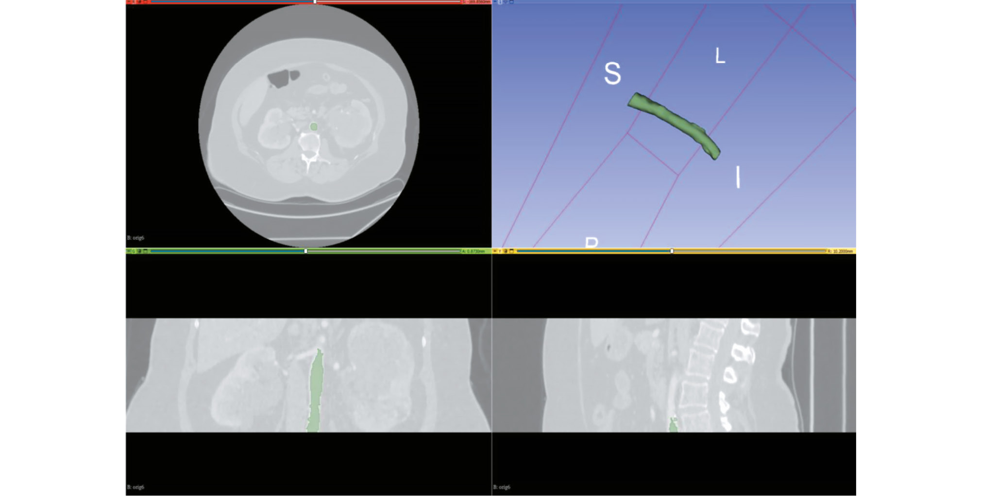
CT angiography dataset with abdominal aorta segmentation
- Authors: Kodenko M.R.1,2, Vasilev Y.A.1, Solovev A.V.1,3, Gatin D.V.1, Yasakova E.P.1, Guseva A.V.1,2, Reshetnikov R.V.1
-
Affiliations:
- Research and Practical Clinical Center for Diagnostics and Telemedicine Technologies
- Bauman Moscow State Technical University
- Morozov Children's City Clinical Hospital
- Issue: Vol 6, No 1 (2025)
- Pages: 23-32
- Section: Datasets
- URL: https://journals.rcsi.science/DD/article/view/310049
- DOI: https://doi.org/10.17816/DD635589
- ID: 310049
Cite item

Abstract
BACKGROUND: Artificial intelligence algorithms are used to analyze images obtained through radiological diagnostic methods. The effectiveness of such algorithms depends on the availability of relevant and representative training datasets. The volume of such data in the public domain should be increased, particularly datasets containing abdominal aorta computed tomography angiography images, with pathology classification and vessel segmentation. The limitations of existing solutions include small sample sizes, restricted dataset specialization, and inconsistent dataset preparation methodologies.
Aim: To create an open dataset containing computed tomography angiography images of abdominal aorta segmentation for normal aorta, aneurysm, thrombosis, and calcification.
MATERIALS AND METHODS: A technical specification for dataset preparation was developed according to the methodology for testing artificial intelligence algorithms, the required sample size was calculated, and approval was obtained from an independent ethics committee. Regarding dataset creation, a previously developed original semiautomatic segmentation algorithm using Slicer 3D software was employed. The inclusion criteria were computed tomography angiography or abdominal computed tomography scans with contrast, arterial phase, and slice thickness ≤3 mm. Conversely, the exclusion criteria were presence of foreign bodies in the aorta lumen and aortic dissection. The algorithm was tested on patient data obtained from the Unified Radiological Information System. An expert evaluation was conducted to assess the compliance of obtained results with the established requirements and evaluate the time efficiency of using the developed segmentation algorithm.
RESULTS: The calculated sample size was 100 angiographic studies, including arterial phase scans with a slice thickness of ≤1.2 mm. Population data: number of unique patients, 100; percentage of female patients, 51%; and median age, 62 years (age range: 18–84 years). Pathology (including combined pathology) was detected in 61% of cases: 60 studies showed signs of calcification, 18 revealed aortic dilation, and 18 determined signs of thrombosed lumen. The average time to process one study (100 slices) using the developed segmentation algorithm was 0.8 hours.
CONCLUSIONS: A dataset containing 100 computed tomography angiography results with abdominal aorta segmentation for normal cases, aneurysm, thrombosis, and calcification was created. The dataset is publicly available and can be used for developing and testing artificial intelligence algorithms and for anthropomorphic modeling of the abdominal aorta.
Full Text
##article.viewOnOriginalSite##About the authors
Maria R. Kodenko
Research and Practical Clinical Center for Diagnostics and Telemedicine Technologies; Bauman Moscow State Technical University
Author for correspondence.
Email: m.r.kodenko@yandex.ru
ORCID iD: 0000-0002-0166-3768
SPIN-code: 5789-0319
Cand. Sci. (Engineering)
Russian Federation, Moscow; MoscowYuriy A. Vasilev
Research and Practical Clinical Center for Diagnostics and Telemedicine Technologies
Email: VasilevYA1@zdrav.mos.ru
ORCID iD: 0000-0002-5283-5961
SPIN-code: 4458-5608
MD, Cand. Sci. (Medicine)
Russian Federation, MoscowAlexander V. Solovev
Research and Practical Clinical Center for Diagnostics and Telemedicine Technologies; Morozov Children's City Clinical Hospital
Email: SolovevAV10@zdrav.mos.ru
ORCID iD: 0000-0003-4485-2638
SPIN-code: 9654-4005
Russian Federation, Moscow; Moscow
Denis V. Gatin
Research and Practical Clinical Center for Diagnostics and Telemedicine Technologies
Email: GatinDV@zdrav.mos.ru
ORCID iD: 0000-0002-6218-3012
SPIN-code: 2256-3564
Russian Federation, Moscow
Elena P. Yasakova
Research and Practical Clinical Center for Diagnostics and Telemedicine Technologies
Email: YasakovaEP@zdrav.mos.ru
ORCID iD: 0000-0003-0315-5502
SPIN-code: 1047-4692
MD, Cand. Sci. (Medicine)
Russian Federation, MoscowAnastasia V. Guseva
Research and Practical Clinical Center for Diagnostics and Telemedicine Technologies; Bauman Moscow State Technical University
Email: GusevaAV13@zdrav.mos.ru
ORCID iD: 0009-0006-1787-4726
SPIN-code: 2778-3820
Russian Federation, Moscow; Moscow
Roman V. Reshetnikov
Research and Practical Clinical Center for Diagnostics and Telemedicine Technologies
Email: ReshetnikovRV1@zdrav.mos.ru
ORCID iD: 0000-0002-9661-0254
SPIN-code: 8592-0558
Cand. Sci. (Physics and Mathematics)
Russian Federation, MoscowReferences
- Kumar DS, Bhat V, Gadabanahalli K, Kalyanpur A. Spectrum of abdominal aortic disease in a tertiary health care setup: MDCT based observational study. J Clin Diagn Res. 2016;10(11):TC24–TC29. doi: 10.7860/JCDR/2016/21373.8928
- Russian Society of Angiologists and Vascular Surgeons. Abdominal aortic aneurysm: clinical guidelines [Internet]. Moscow: Russian Society of Angiologists and Vascular Surgeons; 2022 [cited 2022 Apr 7]. (In Russ.) Available from: https://angiolsurgery.org/library/recommendations/2022/aneurysm/recommendation.pdf
- Baliyan V, Shaqdan K, Hedgire S, Ghoshhajra B. Vascular computed tomography angiography technique and indications. Cardiovascular Diagnosis and Therapy. 2019;9(S1):S14S27. doi: 10.21037/CDT.2019.07.04 EDN: IPZHHC
- Alowais ShA, Alghamdi SS, Alsuhebany N, et al. Revolutionizing healthcare: the role of artificial intelligence in clinical practice. BMC Medical Education. 2023;23(1):689. doi: 10.1186/s12909-023-04698-z EDN: AJSDXW
- Ueda D, Kakinuma T, Fujita SH, et al. Fairness of artificial intelligence in healthcare: review and recommendations. Japanese Journal of Radiology. 2023;42(1):3–15. doi: 10.1007/s11604-023-01474-3 EDN: WQQDIA
- Shchupakova AN, Litvyakov AM. Characteristics of atherosclerotic lesion of the abdominal aorta and its unpaired visceral branches in patients with chronic abdominal ischemia. Terapevticheskii arkhiv. 2004;79(6):70–74. EDN: OJZUCJ
- Radl L, Jin YU, Pepe A, et al. AVT: Multicenter aortic vessel tree CTA dataset collection with ground truth segmentation masks. Data in Brief. 2022;40:107801. doi: 10.1016/j.dib.2022.107801 EDN: PEOYKJ
- Imran M, Kreds JR, Gopu VRR, et al. CIS-UNet: Multi-class segmentation of the aorta in computed tomography angiography via context-aware shifted window self-attention. Computerized Medical Imaging and Graphics. 2024;118:102470. doi: 10.1016/j.compmedimag.2024.102470
- Fantazzini A, Esposito M, Finotello A, et al. 3D automatic segmentation of aortic computed tomography angiography combining multi-view 2D convolutional neural networks. Cardiovascular Engineering and Technology. 2020;11(5):576–586. doi: 10.1007/s13239-020-00481-z EDN: FHKUXK
- Jung Y, Kim S, Kim J, et al. Abdominal aortic thrombus segmentation in postoperative computed tomography angiography images using Bi-directional cnvolutional long short-term memory architecture. Sensors. 2022;23(1):175. doi: 10.3390/s23010175 EDN: SGCHXK
- Norgeot B, Quer G, Beaulieu-Jones BK, et al. Minimum information about clinical artificial intelligence modeling: the MI-CLAIM checklist. Nature Medicine. 2020;26(9):1320–1324. doi: 10.1038/s41591-020-1041-y EDN: NRQASJ
- Vasilev YuA, Arzamasov KM, Vladzymyrskyy AV, et al. Preparing a dataset for training and testing software based on artificial intelligence technology: a training manual. Moscow: Moscow Center for Diagnostics and Telemedicine; 2023. (In Russ.) EDN: OGKFGM
- Tymkovich MYu, Avruninn OG, Semenets VV. Using DICOM images in medical systems. Technical Electrodynamics. 2012;(thematic issue):178–183. (In Russ.)
- Li X, Morgan P, Ashburner J, et al. The first step for neuroimaging data analysis: DICOM to NIfTI conversion. Journal of Neuroscience Methods. 2016;264:47–56. doi: 10.1016/j.jneumeth.2016.03.001
- Kodenko MR, Vasilev YuA, Vladzymyrskyy AV. Segmentation of arterial vessels based on CT angiography data using 3D Slicer software: Guidelines. Moscow: Moscow Center for Diagnostics and Telemedicine; 2024. (In Russ.) EDN: CYLZQL
- Kodenko MR, Makarova TA. Preparation of abdominal computed tomography data set for patients with abdominal aortic aneurysm. Digital Diagnostics. 2023;4(1S):90–92. doi: 10.17816/DD430355 EDN: SIUWRL
- Riley RD, Snell KIE, Archer L, et al. Evaluation of clinical prediction models (part 3): calculating the sample size required for an external validation study. BMJ. 2024;384:e074821. doi: 10.1136/bmj-2023-074821
- Hazra A. Using the confidence interval confidently. Journal of Thoracic Disease. 2017;9(10):4124–4129. doi: 10.21037/jtd.2017.09.14
- Vasilev YuA, Vladzymyrskyy AV, Arzamasov KM, et al. Computer vision in radiation diagnostics: the first stage of the Moscow experiment. [Internet]. Moscow: Izdatel'skie resheniya; 2022 [cited 2024 Apr 22]. Available from: https://telemedai.ru/biblioteka-dokumentov/kompyuternoe-zrenie-v-luchevoj-diagnostike-pervyj-etap-moskovskogo-eksperimenta
- Certificate of state registration of the database N 2024621990/ 08.05.2024. Byul. N 5. Vasilev YuА, Kodenko МR, Solovev AV, et al. A computed tomography angiography dataset showing calcification, thrombosis, dilation, and aneurysm, and containing segmentation of the lumen and wall of the abdominal aorta. Available from: https://elibrary.ru/download/elibrary_67262583_69739205.PDF (In Russ.) EDN: FOPTBQ
- Guseva AV, Kodenko MR. Anthropomorphic abdominal aortic phantoms for computed tomography angiography. Digital Diagnostics. 2024;5(1S):27–29. doi: 10.17816/DD626820 EDN: BMDJUN
- Lesage D, Angelini ED, Bloch I, Funka-Lea G. A review of 3D vessel lumen segmentation techniques: models, features and extraction schemes. Medical image analysis. 2009;13(6):819–845. doi: 10.1016/j.media.2009.07.011
Supplementary files


