


Modelling motor and non-motor signs of early-stage Parkinson's disease
- Authors: Ivanov M.V.1, Kutukova K.A.1
-
Affiliations:
- Research Center of Neurology
- Issue: Vol 16, No 2 (2022)
- Pages: 50-57
- Section: Original articles
- URL: https://journals.rcsi.science/2075-5473/article/view/124058
- DOI: https://doi.org/10.54101/ACEN.2022.2.6
- ID: 124058
Cite item
Full Text

Abstract
Introduction. As Parkinson's disease (PD) develops, a number of non-motor signs precede motor symptoms, including gastrointestinal tract dysfunction. Modelling early-stage PD to comprehensively assess the pattern of morphofunctional changes in the gastrointestinal tract is important in order to develop methods of early disease diagnosis and more effective treatment of autonomic disturbances that are typical in PD, and to increase the patients' quality of life.
Study aim — to offer a model of early-stage PD through long-term oral administration of small doses of the neurotoxin rotenone to rats, and to study the functional and immunohistochemical changes in the gastrointestinal tract of the experimental animals, as well as changes in the substantia nigra.
Materials and methods. The experiment was conducted in male Wistar rats aged 3.0–3.5 months. The study group rats (n = 10) were given rotenone orally at a dose of 5 mg/kg, as a suspension in a 4% carboxymethyl cellulose solution, every second day for 7 months. The control group rats (n = 10) received only the 4% carboxymethyl cellulose solution.
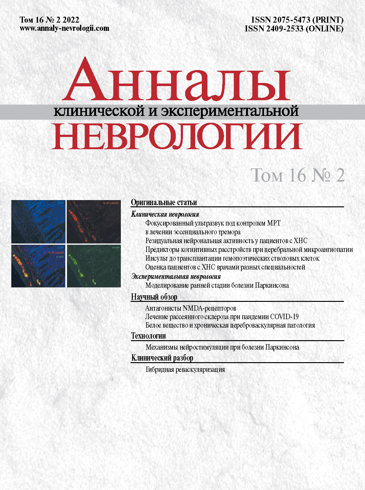
The animals' mobility was assessed at the start and end of the experiment using the open field and narrowing beam-walking test. Gastrointestinal motility was assessed by measuring the passage of dye from the pylorus in a caudal direction along the small intestine. The rats were decapitated and immunohistochemistry was used to assess the density of dopamine neurons in the substantia nigra, nerve fibres, and glia in the Auerbach's plexus of the small intestine, and the location of the total and phosphorylated alpha-synuclein in the enteric nervous system.
Results. Rats in the study group had a statistically significant reduction in the number of dopamine neurons in the substantia nigra. Auerbach's plexus of the small intestine contained significantly less nerve fibres and glia, while fluorescence intensity for alpha-synuclein was increased. Phosphorylated alpha-synuclein was identified in the cholinergic and adrenergic fibres of Auerbach's plexus. Experimental animals had a statistically significant reduction in the gastric emptying rate and small intestine motility compared to the control group.
Conclusion. The presented model of early-stage PD enables the physiological and immunohistochemical symptoms of gastrointestinal dysfunction, similar to that of patients with PD, to be replicated. They are based on intestinal denervation changes and accumulation of abnormal forms of alpha-synuclein in the enteric nervous system.
Full Text
##article.viewOnOriginalSite##About the authors
Mikhail V. Ivanov
Research Center of Neurology
Author for correspondence.
Email: ivanov@neurology.ru
ORCID iD: 0000-0001-5947-9093
junior researcher, Laboratory of neuromorphology
Russian Federation, 125367, Moscow, Volokolamskoye shosse, 80Kristina A. Kutukova
Research Center of Neurology
Email: Chrisbiomag@mail.ru
ORCID iD: 0000-0002-5483-9157
junior researcher, Laboratory of neuromorphology
Russian Federation, 125367, Moscow, Volokolamskoye shosse, 80References
- Klingelhoefer L., Reichmann H. The gut and nonmotor symptoms in Parkinson’s disease. Int. Rev. Neurobiol. 2017; 134: 787–809. doi: 10.1016/bs.irn.2017.05.027
- Pfeiffer R.F. Non-motor symptoms in Parkinson’s disease. Parkinsonism Relat. Disord. 2016;22(Suppl 1): S119–S122. doi: 10.1016/j.parkreldis.2015.09.004
- Jellinger K.A. Synuclein deposition and non-motor symptoms in Parkinson disease. J. Neurol. Sci. 2011; 310(1–2): 107–111. doi: 10.1016/j.jns.2011.04.012
- Braak H., Rub U., Gai W.P., Del Tredici K. Idiopathic Parkinson’s disease: possible routes by which vulnerable neuronal types may be subject to neuroinvasion by an unknown pathogen. J. Neural. Transm. 2003; 110(5): 517–536. doi: 10.1007/s00702-002-0808-2
- Del Tredici K., Rub U., De Vos R.A. et al. Where does Parkinson disease pathology begin in the brain? J. Neuropathol. Exp. Neurol. 2002; 61(5): 413–426. doi: 10.1093/jnen/61.5.413
- Del Tredici K., Braak H. A not entirely benign procedure: progression of Parkinson’s disease. Acta Neuropathol. 2008; 115(4): 379–384. doi: 10.1007/s00401-008-0355-5
- Rietdijk C.D., Perez-Pardo P., Garssen J. et al. Exploring Braak’s hypothesis of Parkinson’s disease. Front. Neurol. 2017; 8: 37. doi: 10.3389/fneur.2017.00037
- Innos J., Hickey M.A. Using rotenone to model Parkinson’s Disease in mice: a review of the role of pharmacokinetics. Chem. Res. Toxicol. 2021; 34(5): 1223–1239. DOI: 1021/acs.chemrestox.0c00522
- Tanner C.M., Kamel F., Ross G.W. et al. Rotenone, paraquat, and Parkinson’s disease. Environ. Health Perspect. 2011; 119(6): 866–872. doi: 10.1289/ehp.1002839
- Betarbet R., Sherer T.B., MacKenzie G. et al. Chronic systemic pesticide exposure reproduces features of Parkinson’s disease. Nat. Neurosci. 2000; 3(12): 1301–136. doi: 10.1038/81834
- Miyazaki I., Isooka N., Imafuku F. et al. Chronic systemic exposure to low-dose rotenone induced central and peripheral neuropathology and motor deficits in mice: reproducible animal model of Parkinson’s disease. Int. J. Mol. Sci. 2020; 21(9): 3254. doi: 10.3390/ijms21093254
- Pan-Montojo F., Anichtchik O., Dening Y. et al. Progression of Parkinson’s disease pathology is reproduced by intragastric administration of rotenone in mice. PLoS One. 2010; 5(1): e8762. doi: 10.1371/journal.pone.0008762
- Pan-Montojo F., Schwarz M., Winkler C. et al. Environmental toxins trigger PD-like progression via increased alpha-synuclein release from enteric neurons in mice. Sci. Rep. 2012; 2: 898. doi: 10.1038/srep00898
- Riederer P., Jellinger K.A., Kolber P. et al. Lateralisation in Parkinson disease. Cell Tissue Res. 2018; 373(1): 297–312. doi: 10.1007/s00441-018-2832-z
- Johnson M.E., Bobrovskaya L. An update on the rotenone models of Parkinson’s disease: their ability to reproduce the features of clinical disease and model gene-environment interactions. Neurotoxicology. 2015; 46: 101–116. doi: 10.1016/j.neuro.2014.12.002
- Dutkiewicz J., Szlufik S., Nieciecki M. et al. Small intestine dysfunction in Parkinson’s disease. J. Neural. Transm. 2015; 122(12): 1659–1661. doi: 10.1007/s00702-015-1442-0
- Marrinan S., Emmanuel A.V., Burn D.J. Delayed gastric emptying in Parkinson’s disease. Mov. Disord. 2014; 29(1): 23–32. doi: 10.1002/mds.25708
- Yan F., Chen Y., Li M. et al. Gastrointestinal nervous system α-synuclein as a potential biomarker of Parkinson disease. Medicine (Baltimore). 2018; 97(28): e11337. doi: 10.1097/MD.0000000000011337
- Иванов М.В., Кутукова К.А., Худоерков Р.М. Морфохимические изменения в нервной системе тонкого кишечника крыс при длительном пероральном введении ротенона. Асимметрия. 2018; 12(4): 217–222. Ivanov M.V., Kutukov K.A., Khudoerkov R.M. Morphochemical changes in the nervous system of the small intestine in rats with prolonged oral administration of rotenone. Journal of asymmetry. 2018; 12(4): 217–222. (In Russ.) doi: 10.18454/ASY.2018.12.4.009
- Phillips R.J., Hudson C.N., Powley T.L. Sympathetic axonopathies and hyperinnervation in the small intestine smooth muscle of aged Fischer 344 rats. Auton. Neurosci. 2013; 179(1–2): 108–121. doi: 10.1016/j.autneu.2013.09.002
- Phillips R.J., Walter G.C., Wilder S.L. et al. Alpha-synuclein-immunopo- sitive myenteric neurons and vagal preganglionic terminals: autonomic pathway implicated in Parkinson’s disease? Neuroscience. 2008; 153: 733–750. doi: 10.1016/j.neuroscience.2008.02.074
- Schmid W., van der Zypen E., Keller H. Die Wirkung einer subtotalen Vago- tomie auf den Plexus myentericus (Auerbach) verschiedener Darmabschnitte. Acta Anat. 1979; 104: 36–51.
- Miyazaki I., Isooka N., Wada K. et al. Effects of enteric environmental modification by coffee components on neurodegeneration in rotenone-treated mice. Cells. 2019; 8(3): 221. doi: 10.3390/cells8030221
- Naudet N., Antier E., Gaillard D. et al. Oral exposure to paraquat triggers earlier expression of phosphorylated α-synuclein in the enteric nervous system of A53T mutant human α-synuclein transgenic mice. J. Neuropathol. Exp. Neurol. 2017; 76(12): 1046–1057. doi: 10.1093/jnen/nlx092
Supplementary files


