


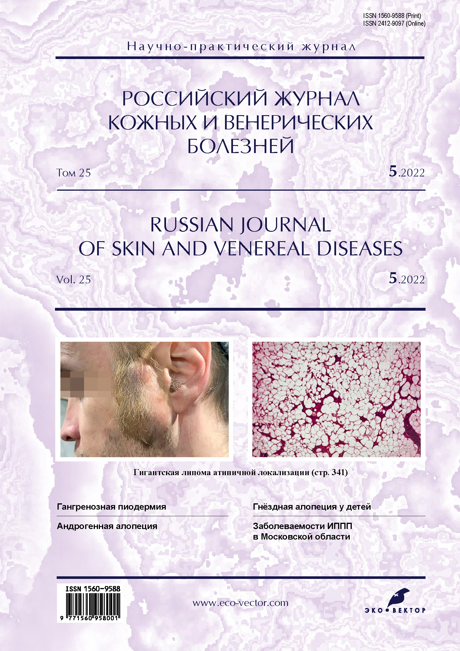
Гигантская липома атипичной локализации
- Авторы: Снарская Е.С.1, Шнахова Л.М.1, Семиклет Ю.М.1
-
Учреждения:
- Первый Московский государственный медицинский университет имени И.М. Сеченова (Сеченовский Университет)
- Выпуск: Том 25, № 5 (2022)
- Страницы: 341-348
- Раздел: ДЕРМАТООНКОЛОГИЯ
- URL: https://journals.rcsi.science/1560-9588/article/view/233225
- DOI: https://doi.org/10.17816/dv112005
- ID: 233225
Цитировать

Аннотация
Липома, или жировая опухоль, жировик (от греч. λίπος ― жир), ― это доброкачественная опухоль мезенхимального происхождения, состоящая из зрелых адипоцитов, патогенез и этиология которой остаются до конца не ясными.
Диагноз основывается на клинической картине, выявлении безболезненных, округлых, подвижных масс с характерным мягким, тестообразным ощущением в подкожных тканях, которые можно найти и в более глубоких тканях, таких как межмышечная септа, органы живота, полость рта, внутренний слуховой канал, угол церебеллопонтина и грудная клетка, вследствие чего к обследованию пациента привлекают профильных специалистов для получения результатов ультразвукового исследования и магнитно-резонансной томографии. Кроме того, проводится гистологическое исследование для исключения липосаркомы.
Лечение липомы зависит от локализации, количества, размеров и субъективных симптомов. Липомы иссекают амбулаторным (под местной анестезией) или методом эндоскопии (малокровно, без риска образования заметных рубцов), открытым способом, что даёт возможность предотвратить рецидивирование.
В связи с высокой частотой развития патологии при наличии наследственной предрасположенности необходимы тщательное обследование родственников пациента и сбор семейного анамнеза.
Представляем клиническое наблюдение гигантской липомы у пациента с редким расположением опухоли на коже височно-скуловой области лица.
Ключевые слова
Полный текст
Открыть статью на сайте журналаОб авторах
Елена Сергеевна Снарская
Первый Московский государственный медицинский университет имени И.М. Сеченова (Сеченовский Университет)
Автор, ответственный за переписку.
Email: snarskaya-dok@mail.ru
ORCID iD: 0000-0002-7968-7663
SPIN-код: 3785-7859
Scopus Author ID: 8714450500
д.м.н., профессор
Россия, 119991, Москва, ул. Большая Пироговская, д. 4, стр. 1Лидия Мухамедовна Шнахова
Первый Московский государственный медицинский университет имени И.М. Сеченова (Сеченовский Университет)
Email: lika-slm@mail.ru
ORCID iD: 0000-0003-3000-0987
ассистент
Россия, МоскваЮлия Михайловна Семиклет
Первый Московский государственный медицинский университет имени И.М. Сеченова (Сеченовский Университет)
Email: semiklet.jul@mail.ru
ORCID iD: 0000-0001-7615-3917
клинический ординатор
Россия, МоскваСписок литературы
- Дерматоонкология / под ред. Г.А. Галил-Оглы, В.А. Молочкова, Ю.В. Сергеева. Москва: Медицина для всех, 2005. С. 787–788.
- Charifa A., Azmat C.E., Badri T. Lipoma pathology. StatPearls [Updated 2021 Oct 2]. Treasure Island (FL): StatPearls Publishing, 2021.
- Kolb L., Yarrarapu S.N., Ameer M.A., et al. Lipoma. StatPearls [Updated 2021 Oct 2]. Treasure Island (FL): StatPearls Publishing, 2021.
- Baldino M.E., Koth V.S., Silva D.N., et al. Gardner syndrome with maxillofacial manifestation: A case report // Spec Care Dentist. 2019. Vol. 39, N 1. Р. 65–71. doi: 10.1111/scd.12339
- Vásquez E.L., Guzman R.P., Sánchez H.M., et al. Poliposis adenomatosa familiar: A propósito de 2 casos [Familiar adenomatous polyposis: report of 2 cases] // Rev Gastroenterol Peru. 2018. Vol. 38, N 1. Р. 78–81. Spanish.
- De Souza Batista F.R., Figueira J.A., Lustosa R.M., et al. Lipoma in the face associated with maxillofacial trauma // J Craniofac Surg. 2017. Vol. 28, N 1. Р. 295–296. doi: 10.1097/SCS.0000000000003255
- Aust M.C., Spies M., Kall S., et al. Posttraumatic lipoma: Fact or fiction? // Skinmed. 2007. Vol. 6, N 6. Р. 266–270. doi: 10.1111/j.1540-9740.2007.06361.x
- Бахмутова Е.Е., Аскерова А.Н., Бабаева Д.М., и др. Дифференциальная диагностика жиросодержащих образований забрюшинного пространства // Медицинская визуализация. 2016. № 2. С. 90–99.
- Энциклопедия клинической онкологии: руководство для практикующих врачей / под ред. М.И. Давыдова, Г.Л. Вышковского. Москва: РЛС, 2005. С. 365–366.
- Усольцев Д.М., Давидян А.А., Бабич Р.А. Опыт удаления гигантских липом в условиях центра амбулаторной хирургии // Стационарозамещающие технологии: амбулаторная хирургия. 2016. № 1-2. С. 94–96.
- Salam G.A. Lipoma excision // Am Fam Physician. 2002. Vol. 65, N 5. Р. 901–904.
Дополнительные файлы


