


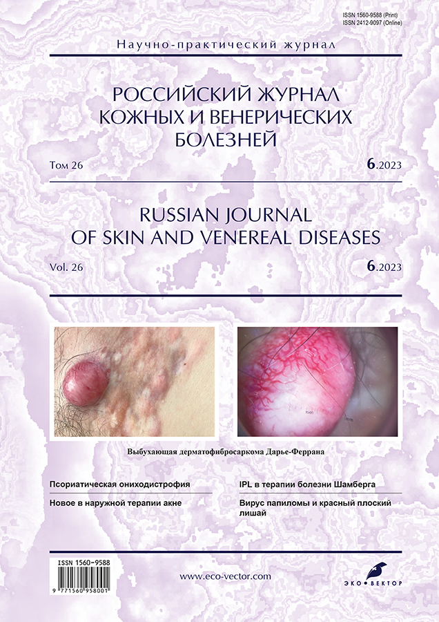
Выбухающая дерматофибросаркома Дарье–Феррана: под маской рубца
- Авторы: Вертиева Е.Ю.1, Каюмова Л.Н.1, Бобкова А.Е.1
-
Учреждения:
- Первый Московский государственный медицинский университет имени И.М. Сеченова (Сеченовский Университет)
- Выпуск: Том 26, № 6 (2023)
- Страницы: 545-552
- Раздел: ДЕРМАТООНКОЛОГИЯ
- URL: https://journals.rcsi.science/1560-9588/article/view/254766
- DOI: https://doi.org/10.17816/dv568542
- ID: 254766
Цитировать

Аннотация
Выбухающая дерматофибросаркома Дарье–Феррана ― редкая мезенхимальная опухоль фиброгистиоцитарного генеза, средней степени злокачественности, с низкой частотой метастазирования, патогенез и этиология которой до конца не ясны. Клиническая картина, на которой основывается диагноз, представлена бляшкой или папулой плотной консистенции красно-коричневого цвета, которая впоследствии постепенно становится болезненным узлом, иногда с изъязвлениями. Гистологическое исследование определяет инфильтрат с муаровым рисунком в дерме в виде веретеновидных клеток с диффузным окрашиванием CD34+ при иммуногистохимическом анализе. Лечение выбухающей дерматофибросаркомы представляет серьёзную проблему в связи с высокой склонностью к рецидивам после удаления. Описанные в литературе методы включают широкое иссечение, операцию по Мосу, химиотерапию, лучевую терапию и таргетное лечение противоопухолевым ингибитором протеинтирозинкиназы.
В работе представлено собственное клиническое наблюдение пациента с выбухающей дерматофибросаркомой Дарье–Феррана, у которого ошибка в постановке диагноза только лишь визуальным методом и неправильно выбранная тактика лечения привели к ухудшению кожного процесса. Интерес данного клинического случая заключается в трудности постановки диагноза при первичном обращении, сложности дифференциальной и морфологической диагностики.
Ключевые слова
Полный текст
Открыть статью на сайте журналаОб авторах
Екатерина Юрьевна Вертиева
Первый Московский государственный медицинский университет имени И.М. Сеченова (Сеченовский Университет)
Email: ivertieva@gmail.com
ORCID iD: 0000-0002-1088-2911
SPIN-код: 3712-8453
кандидат медицинских наук
Россия, 119991, Москва, ул. Трубецкая, д. 8, стр. 2Ляиля Наилевна Каюмова
Первый Московский государственный медицинский университет имени И.М. Сеченова (Сеченовский Университет)
Автор, ответственный за переписку.
Email: avestohka2005@inbox.ru
ORCID iD: 0000-0003-0301-737X
SPIN-код: 4391-9553
кандидат медицинских наук, ассистент
Россия, 119991, Москва, ул. Трубецкая, д. 8, стр. 2Анна Евгеньевна Бобкова
Первый Московский государственный медицинский университет имени И.М. Сеченова (Сеченовский Университет)
Email: anya_bobkova98@mail.ru
ORCID iD: 0000-0003-3611-0917
SPIN-код: 5345-5746
Россия, 119991, Москва, ул. Трубецкая, д. 8, стр. 2
Список литературы
- Клинические рекомендации [интернет]. Саркомы мягких тканей. 2022-2023-2024 (25.07.2022). Утверждены Минздравом РФ. Режим доступа: http://disuria.ru/_ld/12/1283_kr22C49MZ.pdf. Дата обращения: 15.10.2023.
- Ващенко Л.Н., Дашкова И.Р., Салатов Р.Н., и др. К вопросу о хирургическом лечении сарком мягких тканей // Международный журнал прикладных и фундаментальных исследований. 2015. № 8. C. 234–238.
- Brooks J., Ramsey M.L. Dermatofibrosarcoma protuberans. In: StatPearls [Internet]. Treasure Island (FL): StatPearls Publishing, 2023.
- Чупров И.Н., Сыдиков А.А., Заславский Д.В., Насыров Р.А. Дерматоонкопатология: иллюстрированное руководство для врачей / под ред. И.Н. Чупрова, А.А. Сыдикова. Москва: ГЭОТАР-Медиа, 2021. 528 с.
- Cabral R., Wilford M., Ramdass M.J., et al. Dermatofibrosarcoma protuberans associated with trauma: A case report // Mol Clin Oncol. 2020. Vol. 13, N 5. Р. 51. doi: 10.3892/mco.2020.2121
- De Antoni E., Brambullo T., Pescarini E., et al. Dermatofibrosarcoma protuberans on tattooed skin: A case report // Adv Skin Wound Care. 2020. Vol. 33, N 2. Р. 104–108. doi: 10.1097/01.ASW.0000613548.11947.b4
- Hayakawa K., Matsumoto S., Ae K., et al. Risk factors for distant metastasis of dermatofibrosarcoma protuberans // J Orthop Traumatol. 2016. Vol. 17, N 3. Р. 261–266. doi: 10.1007/s10195-016-0415-x
- Abbott J.J., Oliveira A.M., Nascimento A.G. The prognostic significance of fibrosarcomatous transformation in dermatofibrosarcoma protuberans // Am J Surg Pathol. 2006. Vol. 30, N 4. Р. 436–443. doi: 10.1097/00000478-200604000-00002
- Kibbi N., Wang D., Wang W.L., et al. Dermatofibrosarcoma protuberans in pregnancy: A case series and review of the literature // Int J Dermatol. 2021. Vol. 60, N 9. Р. 1114–1119. doi: 10.1111/ijd.15497
- Parlette L.E., Smith C.K., Germain L.M., et al. Accelerated growth of dermatofibrosarcoma protuberans during pregnancy // J Am Acad Dermatol. 1999. Vol. 41, N 5, Pt. 1. Р. 778–783. doi: 10.1016/s0190-9622(99)70023-x
- Ugurel S., Kortmann R.D., Mohr P. S1 guidelines for dermatofibrosarcoma protuberans (DFSP): Update 2018 // J Dtsch Dermatol Ges. 2019. Vol. 17, N 6. Р. 663–668. doi: 10.1111/ddg.13849
- Lyu A., Wang Q. Dermatofibrosarcoma protuberans: A clinical analysis // Oncol Lett. 2018. Vol. 16, N 2. Р. 1855–1862. doi: 10.3892/ol.2018.8802
- Катина М.А., Лесничая О.В., Рязанова Н.В. Дерматофибросаркома выбухающая в дерматологической практике. Клинический случай // Consilium Medicum. 2022. Т. 24, № 8. C. 523–528. doi: 10.26442/20751753.2022.8.201721
- Hao Х., Billings S.D., Wu F., et al. Dermatofibrosarcoma protuberans: Update on the diagnosis and treatment // J Clin Med. 2020. Vol. 9, N 1752. Р. 22. doi: 10.3390/jcm9061752
- Rutkowski P., Debiec-Rychter M. Current treatment options for dermatofibrosarcoma protuberans // Expert Rev Anticancer Ther. 2015. Vol. 15, N 8. Р. 901–909. doi: 10.1586/14737140.2015.1052799
- Иллюстрированное руководство по дерматологии для подготовки врачей к аккредитации / под ред. О.Ю. Олисовой, Н.П. Теплюк. Москва: ГЭОТАР-Медиа, 2023. 376 с.
- Асанов А.Ю., Филиппова М.Г. Нейрофиброматоз: современное состояние проблемы // Российский журнал кожных и венерических болезней. 2011. № 5. C. 14–20.
- Acosta A.E., Vélez C.S. Dermatofibrosarcoma protuberans // Curr Treat Options Oncol. 2017. Vol. 18, N 9. Р. 56. doi: 10.1007/s11864-017-0498-5
- Mullen J.T. Dermatofibrosarcoma protuberans: Wide local excision versus mohs micrographic surgery // Surg Oncol Clin N Am. 2016. Vol. 25, N 4. Р. 827–839. doi: 10.1016/j.soc.2016.05.011
- Vitiello G.A., Lee A.Y., Berman R.S. Dermatofibrosarcoma protuberans: What is this? // Surg Clin North Am. 2022. Vol. 102, N 4. Р. 657–665. doi: 10.1016/j.suc.2022.05.004
- Mareş T., Răducu L., Avino A., et al. Dermatofibrosarcoma protuberans: One centre experience // Chirurgia (Bucur). 2022. Vol. 117, N 5. Р. 601–607. doi: 10.21614/chirurgia.2778
- Гальчина Ю.С., Гогия Б.Ш., Степанова Ю.А., и др. Дерматофибросаркома: обзор литературы и клиническое наблюдение // Медицинская визуализация. 2016. № 2. C. 119–130.
- Kilian K.J., Ruzicka T., Flaig M., et al. Recurrent fibrosarcomatous dermatofibrosarcoma protuberans. Ultrasound imaging // Hautarzt. 2013. Vol. 64, N 7. Р. 512–515. doi: 10.1007/s00105-013-2559-4
Дополнительные файлы


