

")


人工智能软件的技术缺陷
- 作者: Zinchenko V.V.1, Arzamasov K.M.1, Kremneva E.I.1, Vladzymyrskyy A.V.1, Vasilev Y.A.1
-
隶属关系:
- Scientific and Practical Clinical Center for Diagnostics and Telemedicine Technologies
- 期: 卷 4, 编号 4 (2023)
- 页面: 593-604
- 栏目: 技术说明
- URL: https://journals.rcsi.science/DD/article/view/262976
- DOI: https://doi.org/10.17816/DD501759
- ID: 262976
如何引用文章

详细
论证。人工智能软件性能方面的技术缺陷是确定人工智能软件实用性和临床价值的关键。
该研究的目的是对医学影像分析人工智能软件运行中的技术缺陷进行分析并使之系统化。
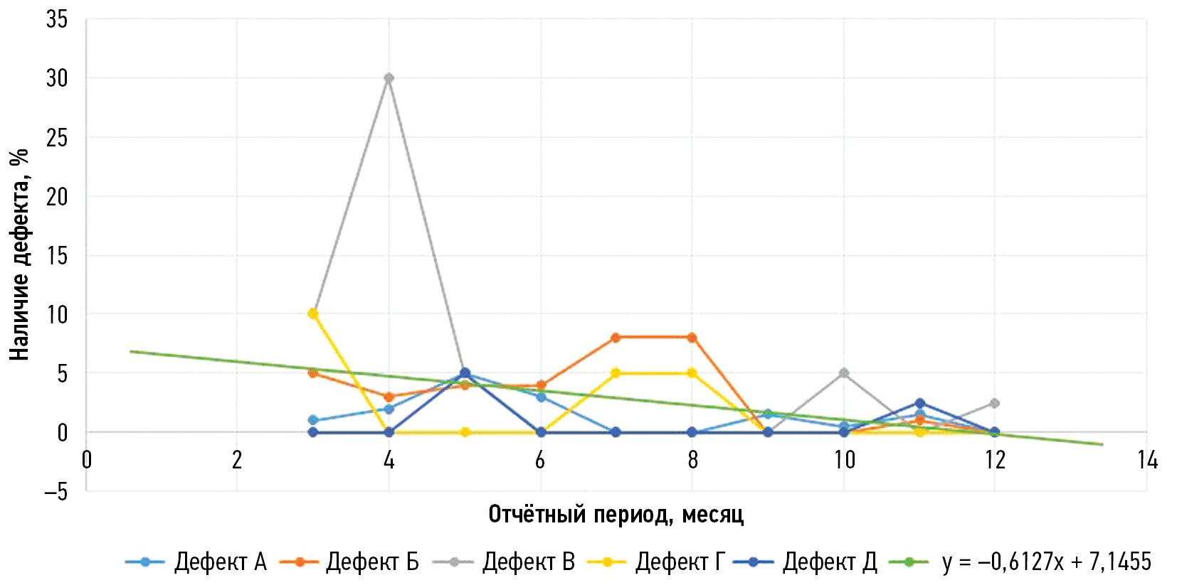
材料和方法。在莫斯科市进行了一项《使用创新计算机视觉技术进行医学图像分析并进一步应用于莫斯科市医疗系统的实验》。在实验框架内,对所有参与解决方案的技术参数进行监测。监测是在批准阶段和试运行阶段进行的。本文以图表形式介绍2021年“乳房摄影术”预防方向的平均技术缺陷数量。这一时期被选为最有意义的时期。这一时期的特点是从提高操作技术稳定性的角度出发,积极开发人工智能软件。为了评估该方法在发现技术缺陷方面的适用性,我们对2022-2023年脑部CT扫描颅内出血的检测方向进行了类似的分析。
结果。本研究分析了“乳房摄影术”(2种算法)和“脑计算机断层扫描”(1种)模式的人工智能软件。在“乳房X射线照相术”模式中,共收集了14个样本,共有20项研 究。在“脑计算机断层扫描”模式中,共收集了12个样本,共有80项研究。我们对每种缺陷类型都绘制了图表,对每种模式绘制了趋势线。趋势线公式的系数表明了,技术缺陷的数量呈下降趋势。
结论。通过分析,我们发现了减少技术缺陷数量的趋势。这可能表明人工智能软件的完善,以及通过定期监测,软件质量的提升。此外,这一结果还显示使用预防和应急方法的通用性。
作者简介
Viktoria V. Zinchenko
Scientific and Practical Clinical Center for Diagnostics and Telemedicine Technologies
编辑信件的主要联系方式.
Email: ZinchenkoVV1@zdrav.mos.ru
ORCID iD: 0000-0002-2307-725X
SPIN 代码: 4188-0635
俄罗斯联邦, Moscow
Kirill M. Arzamasov
Scientific and Practical Clinical Center for Diagnostics and Telemedicine Technologies
Email: ArzamasovKM@zdrav.mos.ru
ORCID iD: 0000-0001-7786-0349
SPIN 代码: 3160-8062
MD, Cand. Sci. (Med.)
俄罗斯联邦, MoscowElena I. Kremneva
Scientific and Practical Clinical Center for Diagnostics and Telemedicine Technologies
Email: KremnevaEI@zdrav.mos.ru
ORCID iD: 0000-0001-9396-6063
SPIN 代码: 8799-8092
MD, Cand. Sci. (Med.)
俄罗斯联邦, MoscowAnton V. Vladzymyrskyy
Scientific and Practical Clinical Center for Diagnostics and Telemedicine Technologies
Email: VladzimirskijAV@zdrav.mos.ru
ORCID iD: 0000-0002-2990-7736
SPIN 代码: 3602-7120
MD, Dr. Sci. (Med.)
俄罗斯联邦, MoscowYuriy A. Vasilev
Scientific and Practical Clinical Center for Diagnostics and Telemedicine Technologies
Email: VasilevYA1@zdrav.mos.ru
ORCID iD: 0000-0002-0208-5218
SPIN 代码: 4458-5608
MD, Cand. Sci. (Med.)
俄罗斯联邦, Moscow参考
- Vladzimirskii AV, Vasil’ev YuA, Arzamasov KM, et al. Computer Vision in Radiologic Diagnostics: the First Stage of Moscow experiment. Vasil’ev YuA, Vladzimirskii AV, editors. Publishing solutions; 2022. (In Russ).
- Ranschaert ER, Morozov S, Algra PR, editors. Artificial Intelligence in Medical Imaging. Berlin: Springer; 2019. doi: 10.1007/978-3-319-94878-2
- Gusev AV, Dobridnyuk SL. Artificial intelligence in medicine and healthcare. Information Society Journal. 2017;(4-5):78–93. (In Russ).
- Shutov DV, Sharova DE, Abuladze LR, Drozdov DV. Artificial intelligence in clinical physiology: How to improve learning agility. Digital Diagnostics. 2023;4(1):81–88. doi: 10.17816/DD123559
- Meldo AA, Utkin LV, Trofimova TN. Artificial intelligence in medicine: current state and main directions of development of the intellectual diagnostics. Diagnostic radiology and radiotherapy. 2020;11(1):9–17. doi: 10.22328/2079-5343-2020-11-1-9-17
- Recht MP, Dewey M, Dreyer K, et al. Integrating artificial intelligence into the clinical practice of radiology: challenges and recommendations. European radiology. 2020;30(6):3576–3584. doi: 10.1007/s00330-020-06672-5
- Larson DB, Harvey H, Rubin DL, et al. Regulatory Frameworks for Development and Evaluation of Artificial Intelligence-Based Diagnostic Imaging Algorithms: Summary and Recommendations. Journal of the American College of Radiology. 2021;18(3 Pt A):413–424. doi: 10.1016/j.jacr.2020.09.060
- Zinchenko V, Chetverikov S, Ahmad E, et al. Changes in software as a medical device based on artificial intelligence technologies. International Journal of Computer Assisted Radiology and Surgery. 2022;17:1969–1977. doi: 10.1007/s11548-022-02669-1
- Nomura Y, Miki S, Hayashi N, et al. Novel platform for development, training, and validation of computer-assisted detection/diagnosis software. International Journal of Computer Assisted Radiology and Surgery. 2020;15(4):661–672. doi: 10.1007/s11548-020-02132-z
- Methodological recommendations on the procedure for expert examination of quality, efficiency and safety of medical devices (in terms of software) for state registration under the national system FGBU «VNIIIMT» Roszdravnadzor. Moscow; 2021. (In Russ).
- Pemberton HG, Zaki LAM, Goodkin O, et al. Technical and clinical validation of commercial automated volumetric MRI tools for dementia diagnosis — a systematic review. Neuroradiology. 2021;63:1773–1789. doi: 10.1007/s00234-021-02746-3
- Order of the Moscow City Health Department No. 51 dated 26.01.2021 «On approval of the procedure and conditions for conducting an experiment on the use of innovative technologies in the field of computer vision to analyze medical images and further application in the health care system of the city of Moscow in 2021». (In Russ).
- Chetverikov SF, Arzamasov KM, Andreichenko AE, et al. Approaches to Sampling for Quality Control of Artificial Intelligence in Biomedical Research. Modern Technologies in Medicine. 2023;15(2):19. doi: 10.17691/stm2023.15.2.02
- Zinchenko VV, Arzamasov KM, Chetverikov SF, et al. Methodology for Conducting Post-Marketing Surveillance of Software as a Medical Device Based on Artificial Intelligence Technologies. Modern Technologies in Medicine. 2022;14(5):15–25. doi: 10.17691/stm2022.14.5.02
- Altman DG. Statistics and ethics in medical research: III How large a sample? British medical journal. 1980;281(6251):1336. doi: 10.1136/bmj.281.6251.1336
- Tyrov IA, Vasilyev YuA, Arzamasov KM, et al. Assessment of the maturity of artificial intelligence technologies for healthcare: methodology and its application based on the use of innovative computer vision technologies for medical image analysis and subsequent applicability in the healthcare system of Moscow. Medical Doctor and IT. 2022;(4):76–92. doi: 10.25881/18110193_2022_4_76
- Vladzimirsky AV, Gusev AV, Sharova DE, et al. Health Information System Maturity Assessment Methodology. Medical Doctor and IT. 2022;(3):68–84. doi: 10.25881/18110193_2022_3_68
- Order of the Moscow City Health Department No. 160 dated 03.11.2022 «On Approval of the Procedure and Conditions for Conducting an Experiment on the Use of Innovative Technologies in Computer Vision for Analyzing Medical Images and Further Application in the Moscow City Health Care System in 2022». (In Russ).
补充文件


