

")


Мальформация Абернети: клинический случай
- Авторы: Панюкова А.В.1, Синицын В.Е.1, Мершина Е.А.1, Ручьёва Н.А.2
-
Учреждения:
- Московский государственный университет имени М.В. Ломоносова, Медицинский научно-образовательный центр
- Национальный медицинский исследовательский центр трансплантологии и искусственных органов имени академика В.И. Шумакова
- Выпуск: Том 4, № 2 (2023)
- Страницы: 226-237
- Раздел: Клинические случаи и серии клинических случаев
- URL: https://journals.rcsi.science/DD/article/view/146888
- DOI: https://doi.org/10.17816/DD289714
- ID: 146888
Цитировать

Аннотация
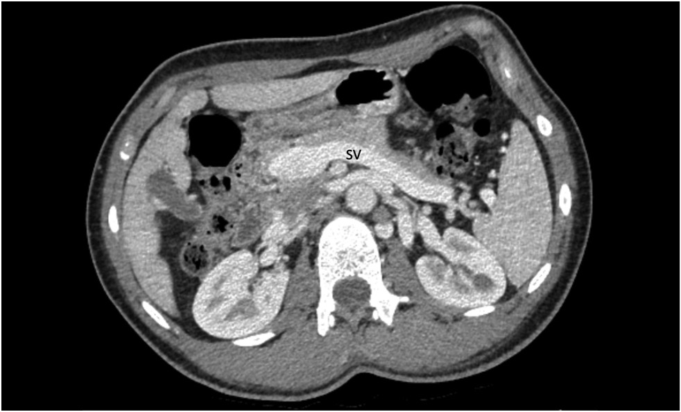
В статье описан клинический случай мальформации Абернети типа Ib у 15-летнего пациента с длительным анамнезом повышенного артериального давления, рецидивирующими носовыми кровотечениями, болью в груди, головокружением, одышкой, низкой толерантностью к физической нагрузке, эпизодами крови в стуле, болью в эпигастральной области, тошнотой и зудом. В результате проведённого комплексного обследования у пациента была диагностирована аномалия развития портальной системы: расширенный кондуит воротной вены, впадающий непосредственно в нижнюю полую вену. Выявлены также множественные узлы в паренхиме печени, расширение камер сердца, гипертрофия миокарда и лёгочная гипертензия. Учитывая выраженность симптомов, размеры и тип шунта, междисциплинарным консилиумом рекомендована трансплантация печени.
В статье рассматриваются алгоритмы диагностики и другие возможные варианты лечения аномалий развития портальной системы.
Полный текст
Открыть статью на сайте журналаОб авторах
Александра Вадимовна Панюкова
Московский государственный университет имени М.В. Ломоносова, Медицинский научно-образовательный центр
Email: panyukovaalexandra@gmail.com
ORCID iD: 0000-0002-5367-280X
Россия, Москва
Валентин Евгеньевич Синицын
Московский государственный университет имени М.В. Ломоносова, Медицинский научно-образовательный центр
Email: vsini@mail.ru
ORCID iD: 0000-0002-5649-2193
SPIN-код: 8449-6590
д-р мед. наук, профессор
Россия, МоскваЕлена Александровна Мершина
Московский государственный университет имени М.В. Ломоносова, Медицинский научно-образовательный центр
Email: elena_mershina@mail.ru
ORCID iD: 0000-0002-1266-4926
SPIN-код: 6897-9641
канд. мед. наук, доцент
Россия, МоскваНаталья Александровна Ручьёва
Национальный медицинский исследовательский центр трансплантологии и искусственных органов имени академика В.И. Шумакова
Автор, ответственный за переписку.
Email: rna1969@yandex.ru
ORCID iD: 0000-0002-8063-4462
канд. мед. наук
Россия, МоскваСписок литературы
- Bernard O., Franchi-Abella S., Branchereau S., et al. Congenital portosystemic shunts in children: Recognition, evaluation, and management // Seminars Liver Dis. 2012. Vol. 32, N 4. P. 273–287. doi: 10.1055/s-0032-1329896
- Papamichail M., Pizanias M., Heaton N. Congenital portosystemic venous shunt // Eur J Pediatrics. 2018. Vol. 177, N 3. P. 285–294. doi: 10.1007/s00431-017-3058-x
- Abernethy J. Account of two instances of uncommon formation in the viscera of the human body: From the philosophical transactions of the royal society of London // Med Facts Observations. 1797. N 7. P. 100–108.
- Guérin F., Blanc T., Gauthier F., et al. Congenital portosystemic vascular malformations // Seminars Pediatric Sur. 2012. Vol. 21, N 3. P. 233–244. doi: 10.1053/j.sempedsurg.2012.05.006
- Born M. The ductus venosus // RoFo Fortschritte Gebiet Rontgenstrahlen Bildgebenden Verfahren. 2021. Vol. 193, N 5. P. 521–526. doi: 10.1055/a-1275-0984
- Baller S.E., Reinehr M., Haslinger C., et al. Case report of neonatal ductus venosus atresia // J Neonatal-Perinatal Med. 2021. Vol. 14, N 2. P. 307–312. doi: 10.3233/NPM-190398
- Franchi-Abella S., Branchereau S., Lambert V., et al. Complications of congenital portosystemic shunts in children: Therapeutic options and outcomes // J Pediatric Gastroenterol Nutrition. 2010. Vol. 51, N 3. P. 322–330. doi: 10.1097/MPG.0b013e3181d9cb92
- Morgan G., Superina R. Congenital absence of the portal vein: Two cases and a proposed classification system for portasystemic vascular anomalies // J Pediatric Sur. 1994. Vol. 29, N 9. P. 1239–1241. doi: 10.1016/0022-3468(94)90812-5
- Tang H., Song P., Wang Z., et al. A basic understanding of congenital extrahepatic portosystemic shunt: Incidence, mechanism, complications, diagnosis, and treatment // Intractable Rare Dis Res. 2020. Vol. 9, N 2. P. 64–70. doi: 10.5582/irdr.2020.03005
- Păcurar D., Dijmărescu I., Dijmărescu A.D., et al. A case report on an incidental discovery of congenital portosystemic shunt // Medicine. 2019. Vol. 98, N 31. P. e16679. doi: 10.1097/MD.0000000000016679
- Shah A., Aziz A., Awwad A., et al. Incidental radiological diagnosis of asymptomatic Abernethy malformations: Two case reports // BJR|Case Reports. 2017. Vol. 3, N 1. P. 20150496. doi: 10.1259/bjrcr.20220059
- Yangín-Ergon E., Ermis N., Colak R., et al. Abernethy malformation type 2 and biliary atresia coexistence: A rare cause of infantile liver transplant // Euroasian J Hepatogastroenterol. 2018. Vol. 8, N 2. P. 163–166. doi: 10.5005/jp-journals-10018-1283
- Sahu M.K., Bisoi A.K., Chander N.C., et al. Abernethy syndrome, a rare cause of hypoxemia: A case report // Ann Pediatric Cardiol. 2015. Vol. 8, N 1. P. 64–66. doi: 10.3389/fcvm.2021.784739
- Lux D., Naito A., Harikrishnan S. Congenital extrahepatic portosystemic shunt with progressive myelopathy and encephalopathy // Practical Neurology. 2019. Vol. 19, N 4. P. 368–371. doi: 10.1136/practneurol-2018-002111
- Merola E., Cao M., La Starza S., et al. Portosystemic encephalopathy in an 86-year-old patient: A clinical challenge // Acta Gastro-Enterologica Belgica. 2016. Vol. 79, N 1. P. 58–59.
- Sharma R., Suddle A., Quaglia A., et al. Congenital extrahepatic portosystemic shunt complicated by the development of hepatocellular carcinoma // Hepatobiliary Pancreatic Dis Int. 2015. Vol. 14, N 5. P. 552–557. doi: 10.1016/S1499-3872(15)60418-0
- Lin X., Rao J., Xiang Y., et al. Case report: A rare syncope case caused by abernethy ii and a review of the literature // Front Cardiovascul Med. 2022. Vol. 8. P. 2050. doi: 10.3389/fcvm.2021.784739
- Hasegawa T., Sato T., Ishii T., et al. Oral sodium phenylbutyrate for hyperammonemia associated with congenital portosystemic shunt: A case report // J Pediatric Endocrinology Metabolism. 2021. Vol. 34, N 3. P. 407–410. doi: 10.1515/jpem-2020-0603
- Peček J., Fister P., Homan M. Abernethy syndrome in Slovenian children: Five case reports and review of literature // World J Gastroenterol. 2020. Vol. 26, N 37. P. 5731–5744. doi: 10.3748/wjg.v26.i37.5731
- Pathak A., Agarwal N., Mandliya J., et al. Abernethy malformation: A case report // BMC Pediatrics. 2012. Vol. 12, N 1. P. 1. doi: 10.1186/1471-2431-12-57
- Duarte-Mesquita R., Sousa M., Vilaverde F., Cardoso R. Abernethy malformation: Beware in cases of unexplained hepatic encephalopathy in adults // BJR| Case Reports. 2017. Vol. 4, N 2. P. 20170054. doi: 10.1259/bjrcr.20170054
- Allegritti M., Enrico B., Basile E., et al. Non-cirrhotic extra-hepatic porto-systemic shunt causing adult-onset encephalopathy treated with endovascular closure // Digestive Diseases and Sciences. 2020. Vol. 65, N 4. P. 946–951. doi: 10.1007/s10620-019-06024-4
- Alvi A.A., Pichardo J., Gupta S., et al. An interesting case of congenital intrahepatic porto-hepatic shunt as a cause of unexplained encephalopathy // Cureus. 2020. Vol. 12, N 4. P. e7639. doi: 10.14309/01.ajg.0000598392.71372.f2
- De Vito C., Tyraskis A., Davenport M., et al. Histopathology of livers in patients with congenital portosystemic shunts (Abernethy malformation): A case series of 22 patients // Virchows Archiv. 2019. Vol. 474, N 1. P. 47–57. doi: 10.1007/s00428-018-2464-4
- Lautz T.B., Shah S.A., Superina R.A. Hepatoblastoma in children with congenital portosystemic shunts // J Pediatric Gastroenterology Nutrition. 2016. Vol. 62, N 4. P. 542–545. doi: 10.1097/MPG.0000000000001012
- Correa C., Luengas J.P., Howard S.C., Veintemilla G. Hepatoblastoma and abernethy malformation type I: Case report // J Pediatric Hematology/Oncology. 2017. Vol. 39, N 2. P. e79–e81. doi: 10.1097/MPH.0000000000000650
- Kwapisz L., Wells M.M., Judaibi B. Al Abernethy malformation: Congenital absence of the portal vein // Can J Gastroenterol Hepatol. 2014. Vol. 28, N 11. P. 587–588. doi: 10.1155/2014/675812
- Benedict M., Rodriguez-Davalos M., Emre S., et al. Congenital extrahepatic portosystemic shunt (abernethy malformation type Ib) with associated hepatocellular carcinoma: Case report and literature review // Pediatric Developmental Pathology. 2017. Vol. 20, N 4. P. 354–362. doi: 10.1177/1093526616686458
- Özden İ., Yavru A., Güllüoğlu M., et al. Transplantation for large liver tumors in the setting of abernethy malformation // Experimental Clin Transplantation. 2017. Vol. 15, Suppl. 2. P. 82–85. doi: 10.6002/ect.TOND16.L23
- Lin K. Y., Chen H., Yu L. Pulmonary arterial hypertension caused by congenital extrahepatic portocaval shunt: A case report // BMC Cardiovascular Disorders. 2019. Vol. 19, N 1. P. 1–5. doi: 10.1186/s12872-019-1124-1
- Osorio M.J., Bonow A., Bond G.J., et al. Abernethy malformation complicated by hepatopulmonary syndrome and a liver mass successfully treated by liver transplantation // Pediatric Transplantation. 2011. Vol. 15, N 7. P. 149–151. doi: 10.1111/j.1399-3046.2010.01337.x
- Baiges A., Turon F., Simón-Talero M., et al. Congenital extrahepatic portosystemic shunts (Abernethy malformation): An international observational study // Hepatology. 2020. Vol. 71, N 2. P. 658–669. doi: 10.1002/hep.30817
- Ponziani F.R., Faccia M., Zocco M.A., et al. Congenital extrahepatic portosystemic shunt: Description of four cases and review of the literature // J Ultrasound. 2019. N 22. P. 349–358. doi: 10.1007/s40477-018-0329-y
- Sheth R., Sivakumar K. The Abernethy malformation with inferior caval vein hypoplasia: A tailored technique for transcatheter closure and an insight into embryological perspective // Cardiology Young. 2018. Vol. 28, N 9. P. 1169–1171. doi: 10.1017/S1047951118000884
- Li H., Ma Z., Xie Y., Tian F. Recurrent hyperammonemia after abernethy malformation Type 2 closure: A case report // Ann Hepatol. 2017. Vol. 16, N 3. P. 460–464. doi: 10.5604/01.3001.0009.8603
- Kanazawa H., Nosaka S., Miyazaki O., et al. The classification based on intrahepatic portal system for congenital portosystemic shunts // J Pediatric Sur. 2015. Vol. 50, N 4. P. 688–695. doi: 10.1016/j.jpedsurg.2015.01.009
Дополнительные файлы


