

")


Computer tomography of uro-lymphatic fistulas associated with renal colic
- Authors: Gelezhe P.B.1,2, Goryacheva K.M.3
-
Affiliations:
- Moscow Center for Diagnostics and Telemedicine
- European Medical Center
- The First Sechenov Moscow State Medical University (Sechenov University)
- Issue: Vol 3, No 2 (2022)
- Pages: 149-155
- Section: Case reports
- URL: https://journals.rcsi.science/DD/article/view/106050
- DOI: https://doi.org/10.17816/DD106050
- ID: 106050
Cite item

Abstract
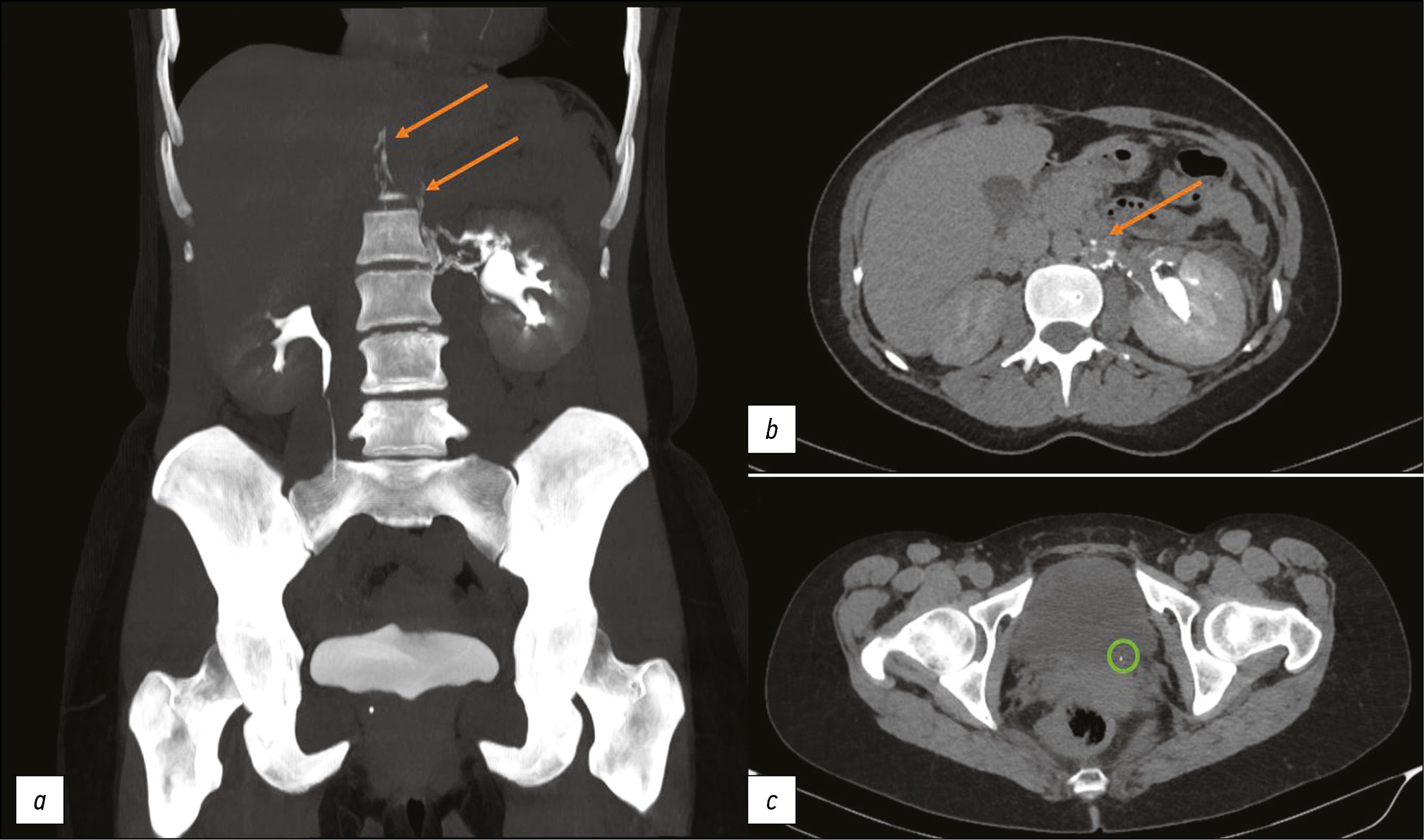
This article presents two clinical observations of uro-lymphatic fistulas diagnosed by computed tomography. In both cases, the patients were admitted with symptoms of renal colic. Uro-lymphatic fistulas are a rare condition caused by the formation of a connection between the urinary and lymphatic systems, which is caused by, as a rule, lymphatic vessel obstruction due to parasitic infestation. Other causes may be radiation therapy, retroperitoneal trauma, and tumor sprouting. In the era before antibiotics, infectious processes such as xanthogranulomatous pyelonephritis and renal tuberculosis were common. Cases of uro-lymphatic fistulas formed against urolithiasis background are presented below. In the clinical cases presented, urine directly entered the lymphatic vessels through a uro-lymphatic fistula detected on contrast-enhanced computed tomography. Uro-lymphatic fistulas caused by impaired urine outflow due to blocked urinary tract are rarely detected since abdominal ultrasound is the diagnostic method of choice in renal colic. In the vast majority of cases, uro-lymphatic fistulas are treated conservatively and do not require surgical intervention. As a rule, the formed fistulas cease to exist when its root cause is successfully treated.
Full Text
##article.viewOnOriginalSite##About the authors
Pavel B. Gelezhe
Moscow Center for Diagnostics and Telemedicine; European Medical Center
Author for correspondence.
Email: gelezhe.pavel@gmail.com
ORCID iD: 0000-0003-1072-2202
SPIN-code: 4841-3234
MD, Cand. Sci. (Med.)
Russian Federation, Moscow; MoscowKristina M. Goryacheva
The First Sechenov Moscow State Medical University (Sechenov University)
Email: cristina.imago27@yandex.ru
ORCID iD: 0000-0003-1221-9694
SPIN-code: 2722-6891
Russian Federation, Moscow
References
- Stainer V, Jones P, Juliebo SO, et al. Chyluria: what does the clinician need to know? Ther Adv Urol. 2020;(12):1756287220940899. doi: 10.1177/1756287220940899
- Roffi F, Eiss D, Petit F, et al. Pyelolymphatic fistula in a patient with lymphatic filariasis: a case report. (In French). J Radiol. 2007;88(12):1896–1898. doi: 10.1016/s0221-0363(07)78369-5
- Yu NC, Raman SS, Patel M, et al. Fistulas of the genitourinary tract: a radiologic review. Radiographics. 2004;24(5):1331–1352. doi: 10.1148/rg.245035219
- McIntosh GH, Morris B. The lymphatics of the kidney and the formation of renal lymph. J Physiology. 1971;214(3):365–376. doi: 10.1113/jphysiol.1971.sp009438
- Skandalakis JE, Skandalakis LJ, Skandalakis PN. Anatomy of the Lymphatics. Surg Oncol Clin N Am. 2007;16(1):1–16. doi: 10.1016/j.soc.2006.10.006
- Diamond E, Schapira HE. Chyluria ― a review of the literature. Urology. 1985;26(5):427–431. doi: 10.1016/0090-4295(85)90147-5
- Rajaonarison N, Ahmad A, Cucchi JM, et al. Reversible uro-lymphatic fistula. Clin Imaging. 2012;36(1):72–74. doi: 10.1016/j.clinimag.2011.04.012
- Graziani G, Cucchiari D, Verdesca S, et al. Chyluria associated with nephrotic-range proteinuria: pathophysiology, clinical picture and therapeutic options. Nephron Clinical Practice. 2011;119(3):248–253. doi: 10.1159/000329154
- Kim RJ, Joudi FN. Chyluria after partial nephrectomy: case report and review of the literature. Sci World J. 2009;(9):1–4. doi: 10.1100/tsw.2009.5
Supplementary files


